

Distribution and localization patterns of estrogen receptor-beta and insulin-like growth factor-1 receptors in neurons and glial cells of the female rat substantia nigra: localization of ERbeta and IGF-1R in substantia nigra
- PMID: 17480015
- PMCID: PMC2907103
- DOI: 10.1002/cne.21358
Distribution and localization patterns of estrogen receptor-beta and insulin-like growth factor-1 receptors in neurons and glial cells of the female rat substantia nigra: localization of ERbeta and IGF-1R in substantia nigra
Abstract
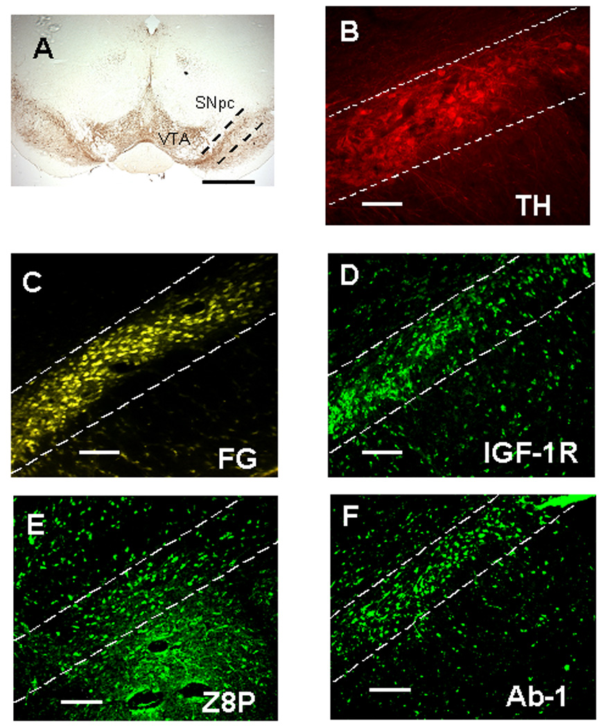
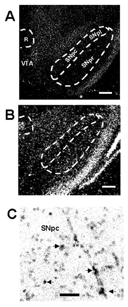
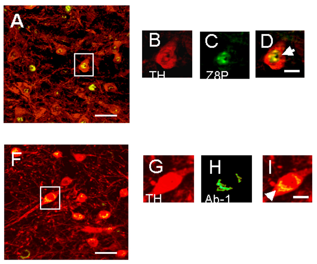
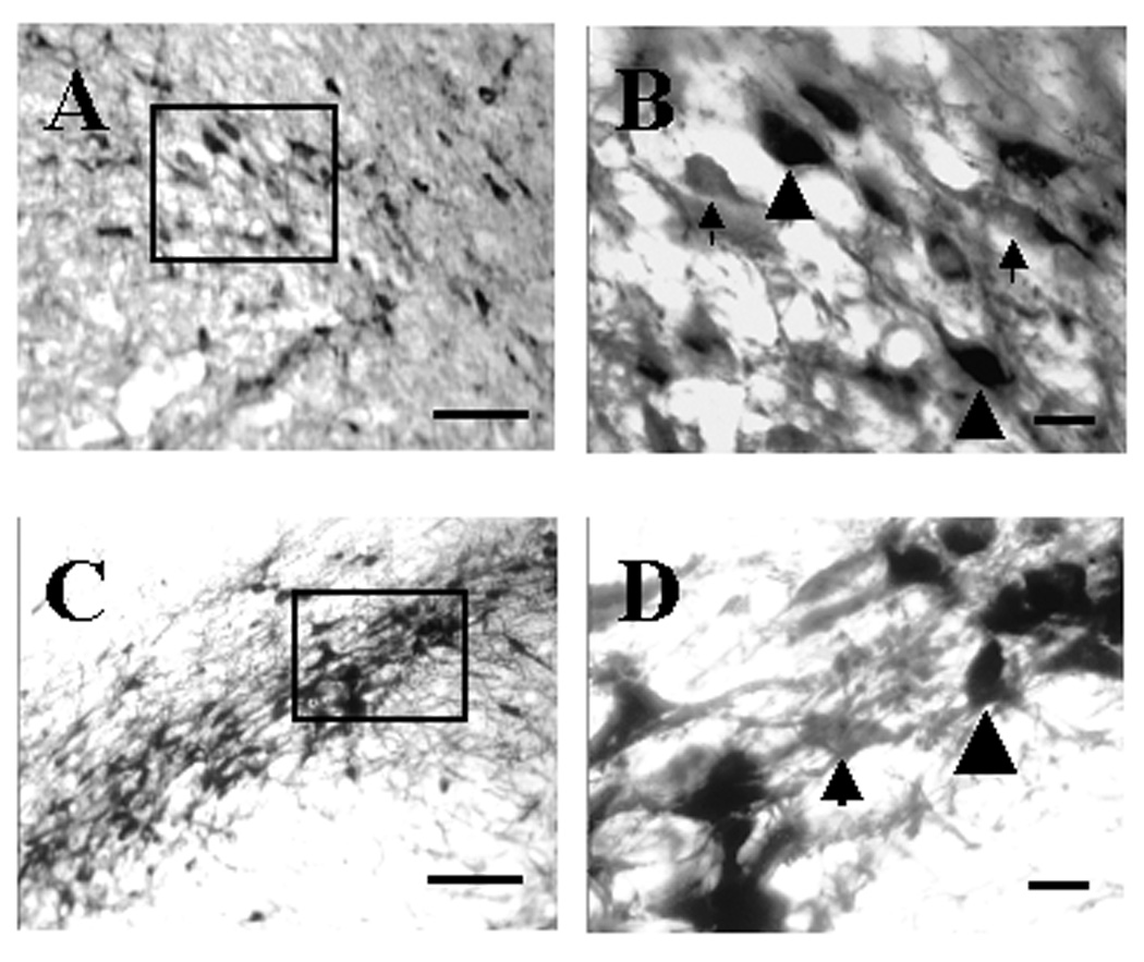
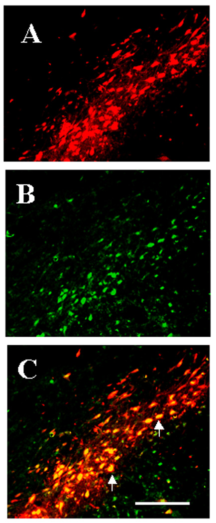
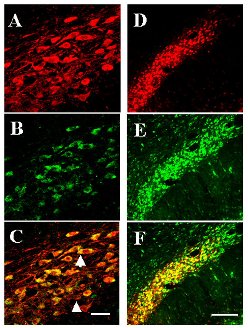
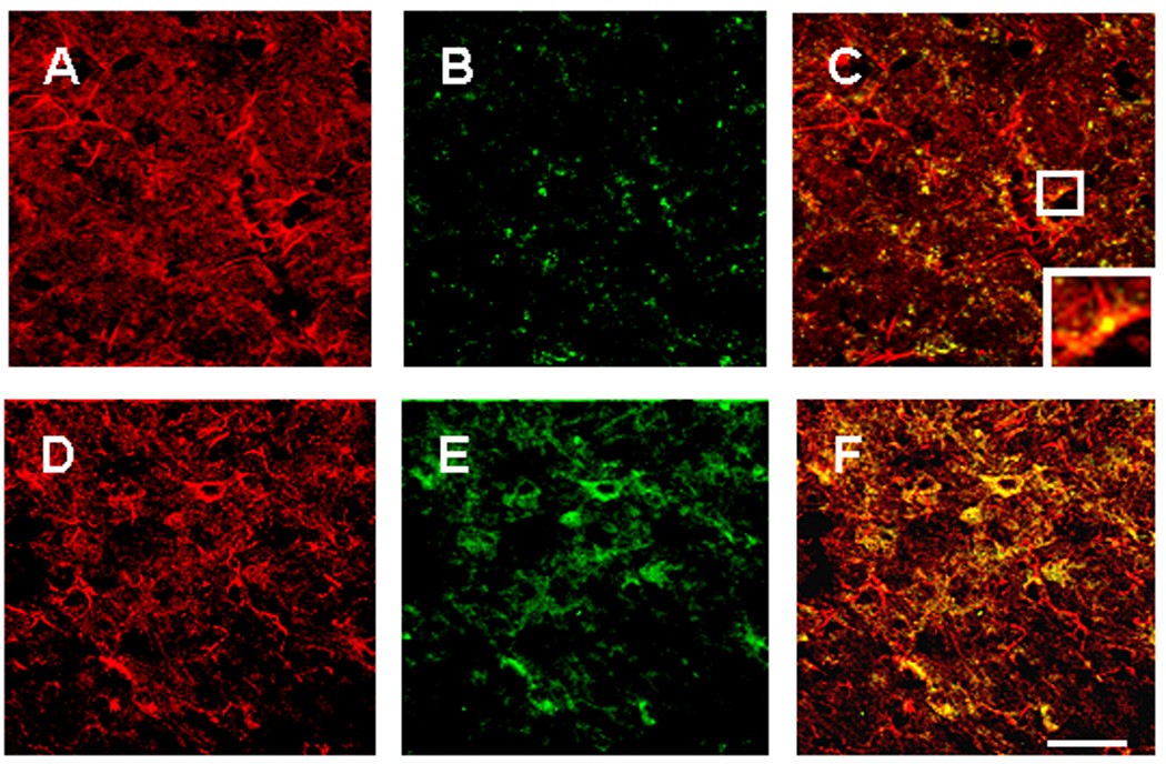
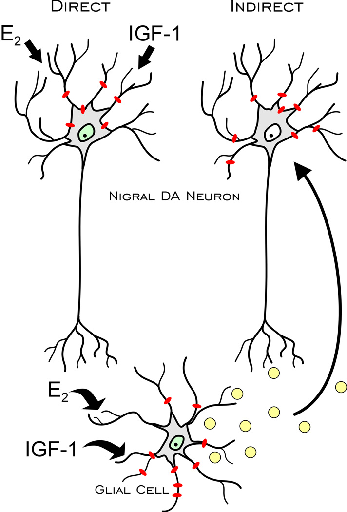
Although several studies have focused on the neuroprotective effects of estrogen (E2) on stroke, there have been tantalizing reports on the potential neuroprotective role of E2 in degenerative neuronal diseases such as Alzheimer's and Parkinson's (PD). In animal models of PD, E2 protects the nigrostriatal dopaminergic (DA) system against neurotoxins. However, little is known about the cellular and molecular mechanism(s) involved by which E2 elicits its neuroprotective effects on the nigrostriatal DA system. A preferred mechanism for neuroprotection is the interaction of E2 with specific neuroprotective growth factors and receptors. One such neuroprotective factor/receptor system is insulin-like growth factor-1 (IGF-1). E2 neuroprotective effects in the substantia nigra (SN) DA system have been shown to be dependent on IGF-1. To determine whether E2 also interacts with the IGF-1 receptor (IGF-1R) and to determine the cellular localization of estrogen receptor (ER) and IGF-1R, we compared the distribution of ER and IGF-1R in the SN. Stereological measurements revealed that 40% of the subpopulation of tyrosine hydroxylase-immunoreactive (TH-ir) SN pars compacta (SNpc) DA neurons are immunoreactive for estrogen receptor-beta (ERbeta). No immunolabeling for ERalpha was observed. In situ hybridization and immunocytochemistry studies confirmed the expression of IGF-1R mRNA and revealed that almost all TH-ir SNpc DA neurons were immunoreactive for IGF-1R, respectively. Moreover, one-third of glial fibrillary acidic protein (GFAP-ir) cells in the SN were ERbeta-ir, and 67% of GFAP-ir cells expressed IGF-1R-ir. Therefore, the localization of ERbeta and IGF-1R on SNpc DA neurons and astrocytes suggests a modulatory role of E2 on IGF-1R, and this modulation may affect neuroprotection.
Figures
References
-
- Azcoitia I, Sierra A, Garcia-Segura LM. Localization of estrogen receptor beta-immunoreactivity in astrocytes of the adult rat brain. Glia. 1999a;26:260–267. - PubMed
-
- Azcoitia I, Sierra A, Garcia-Segura LM. Neuroprotective effects of estradiol in the adult rat hippocampus: interaction with insulin-like growth factor-I signalling. J Neurosci Res. 1999b;58:815–822. - PubMed
-
- Bains M, Cousins JC, Roberts JL. Society for Neuroscience Abstract. Washington D.C: 2005. Estrogen Promotes Neuronal Survival in Mesencephalic Dopamine Progenitor Cultures.
-
- Barrett-Connor E, Bush TL. Estrogen and coronary heart disease in women. Jama. 1991;265:1861–1867. - PubMed
-
- Blurton-Jones M, Tuszynski MH. Estrogen receptor-beta colocalizes extensively with parvalbumin-labeled inhibitory neurons in the cortex, amygdala, basal forebrain, and hippocampal formation of intact and ovariectomized adult rats. J Comp Neurol. 2002;452:276–287. - PubMed
Publication types
MeSH terms
Substances
Grants and funding
LinkOut - more resources
Full Text Sources
Miscellaneous
