. 2007 May 4;3(5):e62.
doi: 10.1371/journal.ppat.0030062.

Impact of Mycobacterium ulcerans biofilm on transmissibility to ecological niches and Buruli ulcer pathogenesis

Affiliations

Impact of Mycobacterium ulcerans biofilm on transmissibility to ecological niches and Buruli ulcer pathogenesis

Laurent Marsollier et al. PLoS Pathog. .

Abstract

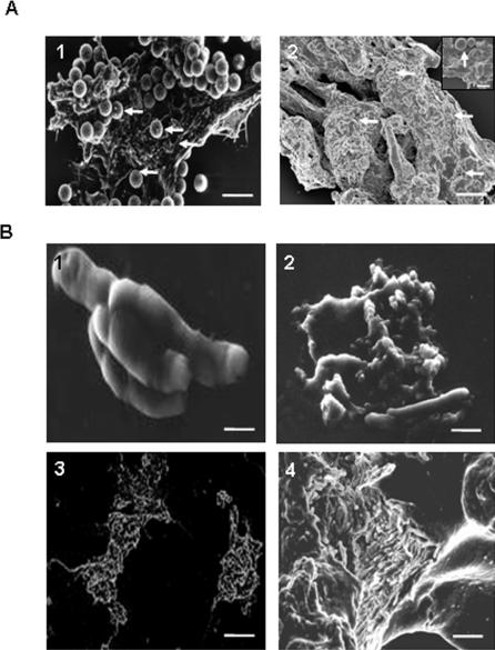
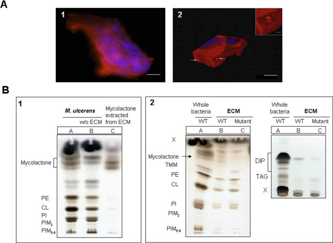
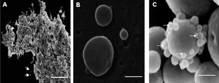
The role of biofilms in the pathogenesis of mycobacterial diseases remains largely unknown. Mycobacterium ulcerans, the etiological agent of Buruli ulcer, a disfiguring disease in humans, adopts a biofilm-like structure in vitro and in vivo, displaying an abundant extracellular matrix (ECM) that harbors vesicles. The composition and structure of the ECM differs from that of the classical matrix found in other bacterial biofilms. More than 80 proteins are present within this extracellular compartment and appear to be involved in stress responses, respiration, and intermediary metabolism. In addition to a large amount of carbohydrates and lipids, ECM is the reservoir of the polyketide toxin mycolactone, the sole virulence factor of M. ulcerans identified to date, and purified vesicles extracted from ECM are highly cytotoxic. ECM confers to the mycobacterium increased resistance to antimicrobial agents, and enhances colonization of insect vectors and mammalian hosts. The results of this study support a model whereby biofilm changes confer selective advantages to M. ulcerans in colonizing various ecological niches successfully, with repercussions for Buruli ulcer pathogenesis.


Conflict of interest statement

Competing interests. The authors have declared that no competing interests exist.

Figures

Figure 1
Figure 1. M. ulcerans ECM and Its Formation
(A) Scanning electron micrographs of bacilli isolated by immunomagnetic beads separation from mammalian host tissues. Bacilli isolated from human (1) or mouse (2) tissue formed large clusters that are surrounded by ECM. Arrows indicate the immunomagnetic beads. Scale bar: 10 μm (1 and 2), 1 μm (inset). (B) Scanning electron micrographs of cluster formation by M. ulcerans. (1) Ten days after inoculation in 7H9 culture medium supplemented with OADC and containing Tween 80 0.05%, some bacilli formed a small cluster. (2) After 20 d, the bacilli had multiplied and the clusters were covered by extracellular material. (3) At the end of exponential growth (45 d after inoculation) a large cluster was formed (4), which was surrounded by the ECM. Scale bars: 1 μm (1), 1.5 μm (2), 25 μm (3), and 10 μm (4).
Figure 2
Figure 2. M. ulcerans ECM Localized Only in the Outermost Bacterial Layer
Transmission electron micrographs of cluster formation by M. ulcerans showed that ECM covers only the peripheral bacterial layer. (A) ECM thickness is estimated to range between 4 and 40 μm (dotted line and arrow). (B) Compared to classical biofilm, the space between M. ulcerans bacteria in the cluster is small (arrows), with little ECM. Scale bars: 1 μm.
Figure 3
Figure 3. Impact of ECM Removal on M. ulcerans Cultivability and Permeability
(A) Scanning electron micrographs of M. ulcerans recovered after ECM extraction. The ECM could be removed by vortexing the cells with glass beads in Tween 80 0.05%. Scale bars: 5 μm. (B) The effect of removal of ECM on the growth of M. ulcerans. CFUs onto Löwenstein–Jensen slants were determined after inoculation of serial dilutions of bacterial suspension (Acid Fast Bacilli [AFB]). Open symbols: CFUs from sample without ECM; filled symbols: bacterial clusters with ECM. Mean value ± standard error of the mean (S.E.M.) of three samples. (C) Potassium release assay in bacterial supernatant of M. ulcerans culture sample taken at different times after mechanical treatment with detergent for ECM removal. Each time point is the mean value ± S.E.M. of three independent assays. Boiled corresponds to the supernatant of boiled bacteria. Significant difference was obtained after 60 s of treatment. (D) Detection of KatG by Western blotting on M. ulcerans culture sample after mechanical treatment with detergent for ECM removal. Bacterial supernatant (30 μl) was taken at different times after treatment by beads and detergent was loaded on a 4%–12% SDS-PAGE gel and transferred to nitrocellulose. Detection was performed with rabbit polyclonal serum anti-KatG serum and anti-rabbit horseradish peroxidise-conjugated IgG. KatG was detected after only 45 s of treatment. Proteins (60 μg) extracted from M. ulcerans lysate were used for positive control.
Figure 4
Figure 4. ECM Is a Poor Source of Antigens
(A) Western blotting with one representative human serum sample on M. ulcerans lysate or on ECM fraction. Proteins (20 μg) extracted from M. ulcerans lysate (lanes 1 and 3) or from ECM (lanes 2 and 4) were loaded on a 4%–12% SDS-PAGE gel and transferred to nitrocellulose. Lanes 1 and 2: Coomassie staining. Lanes 3 and 4: Detection with serum from a Buruli ulcer patient, and anti-human horse radish peroxidase-conjugated IgG. Many antigens were detected in the M. ulcerans lysate, whereas none were found in ECM. (B) Detection of reactive IgG by ELISA in M. ulcerans lysate, membrane, cytosolic, ECM, and vesicle fractions. Samples were divided into three groups according to the patients' clinical stage of disease: no ulceration, limited ulceration, and extensive ulceration. Comparison using one way analysis of variance followed by the Newman–Keuls multiple comparison test shows that the relative titre of IgG binding to antigens from ECM was significantly less compared to that of the other fractions.
Figure 5
Figure 5. Analysis of Carbohydrate and Lipids in ECM
(A) Detection of carbohydrates in M. ulcerans aggregates by multicolor fluorescence microscopy. (1) Bacteria were stained by DAPI, whereas carbohydrates labeled with Texas red hydrazide localized within an ECM. (2) After 3-D construction, channels were observed (arrows and insert) in ECM. Scale bars: 10 μm; insert, 1 μm. (B) TLC analysis of total lipids of M. ulcerans and purification of mycolactone from ECM. (1) The ECM of M. ulcerans wild-type strain was extracted with glass beads and Tween 80. The TLC plate was developed in CHCl3/CH3OH/H20 (65:25:4, vol:vol). Lane A: Total lipids of M. ulcerans with ECM. Lane B: Total lipids of M. ulcerans without ECM. Lane C: The lipids extracted from the ECM were fractionated by adsorption chromatography on a silica gel column and mycolactone was purified by preparative TLC. (2) Lane A: Total lipids extracted from wild-type M. ulcerans before the treatment with glass beads. The ECM of M. ulcerans wild-type (WT) (lane B) and mutant (Mut) strains (lane C) was extracted with glass beads.. The TLC plate was developed in the same solvent system (left) or in petroleum ether/ethyl acetate (98:2, vol:vol, three developments) (right). Lipids were revealed with a cupric sulfate solution with charring. CL, cardiolipin; DIP, phthiodiolone diphthioceranates and phenolphthiodiolone diphthioceranates; PE, phosphatidylethanolamine; PI, phosphatidylinositol; PIMx, phosphatidylinositol mannosides (where x refers to the number of mannose residues); TAG, triacylglycerol; X, unidentified compounds.
Figure 6
Figure 6. Detection and Purification of Vesicles from ECM
(A) Scanning electron micrographs of M. ulcerans cluster showed that the ECM contains vesicles (arrows). Scale bar: 10 μm. (B) Scanning electron micrographs of vesicles purified from a culture supernatant by ultracentrifugation. The vesicle size (50 to 250 nm) is heterogeneous. Scale bar: 200 nm. (C) Scanning electron micrographs of vesicles (arrows) that were purified by immunomagnetic particle separation from an infected mouse tissue. Scale bar: 0.8 μm.
Figure 7
Figure 7. Cytotoxicity of Vesicles
Comparative cytoxicity of vesicles (black bars) and purified mycolactone (grey bars) on phagocytic cells (bone marrow–derived mouse macrophages, [A]) and non-phagocytic cells (Cos cells, [B]). The mycolactone associated with vesicles is more cytotoxic than purified mycolactone.
Figure 8
Figure 8. No M. ulcerans ECM Is Detected in Culture Supplemented with Organic Extracts of Algae or in Salivary Glands of Water Bugs
(A) Scanning electron micrograph of the accessory salivary glands of N. cimicoides showed that the bacilli (wild-type strain) are either alone (yellow arrows) or in clusters (yellow arrowheads) lacking ECM. Scale bar: 10 μm. (B) M. ulcerans aggregate cultured with organic matter extracted from Rhizoclonium sp. The bacilli form large clusters that are not covered by ECM and that adhere to aquatic plant surface (arrow). Scale bar: 20 μm.

References

    1. Marsollier L, Aubry J, Saint-Andre JP, Robert R, Legras P, et al. Ecology and transmission of Mycobacterium ulcerans . Pathol Biol. 2003;51:490–495. - PubMed
    1. Marsollier L, Severin T, Aubry J, Merritt RW, Saint Andre JP, et al. Aquatic snails, passive hosts of Mycobacterium ulcerans . Appl Environ Microbiol. 2004;70:6296–6298. - PMC - PubMed
    1. Marsollier L, Stinear T, Aubry J, Saint Andre JP, Robert R, et al. Aquatic plants stimulate the growth of and biofilm formation by Mycobacterium ulcerans in axenic culture and harbor these bacteria in the environment. Appl Environ Microbiol. 2004;70:1097–1103. - PMC - PubMed
    1. Portaels F, Chemlal K, Elsen P, Johnson PD, Hayman JA, et al. Mycobacterium ulcerans in wild animals. Rev Sci Tech Off int Epiz. 2001;20:252–264. - PubMed
    1. Stinear T, Davies JK, Jenkin GA, Hayman JA, Oppedisano F, et al. Identification of Mycobacterium ulcerans in the environment from regions in southeast Australia in which it is endemic with sequence capture-PCR. Appl Env Microbiol. 2000;66:3206–3213. - PMC - PubMed

Publication types