. 2007 May 3;54(3):403-16.
doi: 10.1016/j.neuron.2007.03.029.

Drosophila sensory neurons require Dscam for dendritic self-avoidance and proper dendritic field organization

Affiliations

Drosophila sensory neurons require Dscam for dendritic self-avoidance and proper dendritic field organization

Peter Soba et al. Neuron. .

Abstract

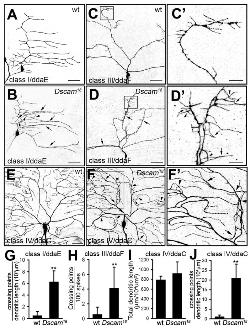
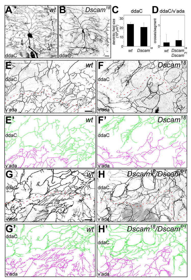
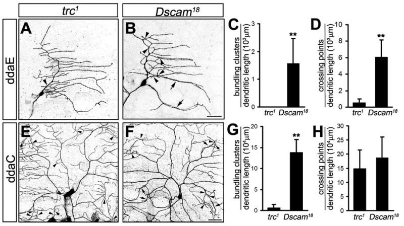
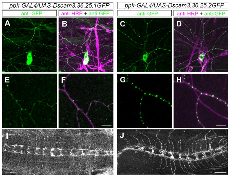
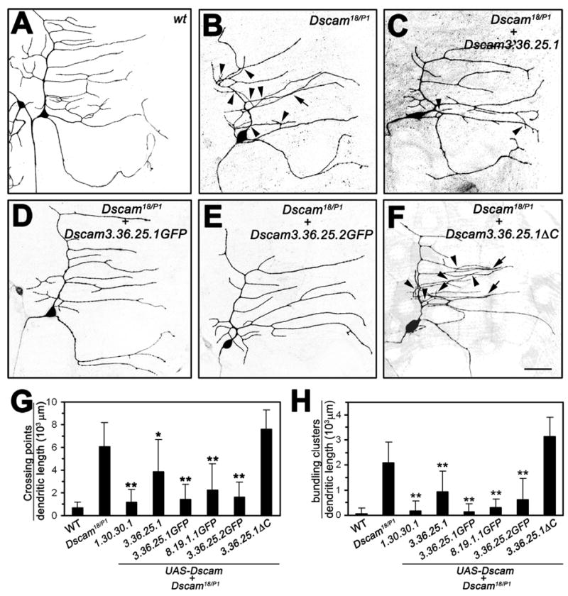
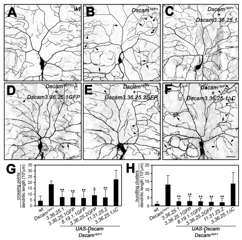
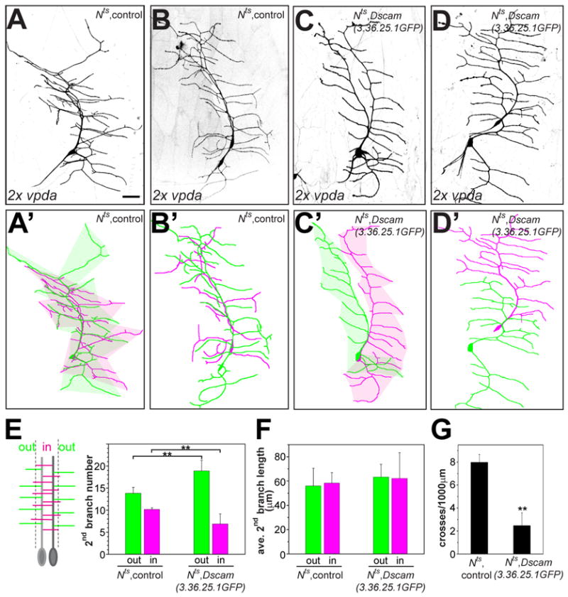
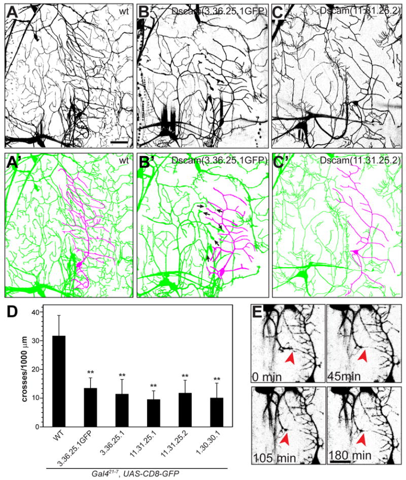
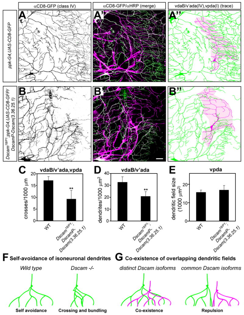
A neuron's dendrites typically do not cross one another. This intrinsic self-avoidance mechanism ensures unambiguous processing of sensory or synaptic inputs. Moreover, some neurons respect the territory of others of the same type, a phenomenon known as tiling. Different types of neurons, however, often have overlapping dendritic fields. We found that Down's syndrome Cell Adhesion Molecule (Dscam) is required for dendritic self-avoidance of all four classes of Drosophila dendritic arborization (da) neurons. However, neighboring mutant class IV da neurons still exhibited tiling, suggesting that self-avoidance and tiling differ in their recognition and repulsion mechanisms. Introducing 1 of the 38,016 Dscam isoforms to da neurons in Dscam mutants was sufficient to significantly restore self-avoidance. Remarkably, expression of a common Dscam isoform in da neurons of different classes prevented their dendrites from sharing the same territory, suggesting that coexistence of dendritic fields of different neuronal classes requires divergent expression of Dscam isoforms.


Figures

Figure 1
Figure 1. MARCM analysis of dendritic morphogenesis of Dscam mutant da neurons
(A–F) Representative MARCM clones of class I ddaE (A–B), class III ddaF (C–D), and class IV ddaC (E–F) neurons. Wild type and Dscam18 mutant MARCM clones are shown as indicated. (C′), (D′), and (F′) are enlarged views of the highlighted areas in (C), (D), and (F). Arrows indicate dendritic bundling and arrowheads indicate crosses of dendritic branches (or spikes in ddaF). Scale bars, 30 μm in (A–F) or 10 μm in (C′, D′, and F′). (G–J) Quantitative analysis of the crossing points of class I ddaE dendrites (G, n=7 for each group), dendritic spikes of ddaF (H, n=7 for each group), total dendritic length (I) and the crossing points of dendritic branches (J) of ddaC neurons (n=6 for each group). All the data are mean ± SD in this and the following figures (**, P<0.01, student’s t-test).
Figure 2
Figure 2. Dscam does not play a major role in interneuronal recognition and tiling
(A–B) MARCM clones of wild type (A) and Dscam18 mutant (B) class IV ddaC neurons. Note that the dendritic field size is largely unchanged in Dscam mutants (B) compared to the wild type (A) and does not show prominent overgrowth. (C) Quantitative analysis of total dendritic field size of wild type (n=4) and Dscam18 (n=5) ddaC MARCM clones. (D) Quantitative analysis of interneuronal dendritic crosses between adjacent ddaC and v’ada class IV da neurons in wild type (n=16) and Dscam18/DscamP1 (n=17) third instar larvae. (E–F) Dendrites of two adjacent MARCM clones of wild type (E and E′) and Dscam18 mutant (F and F′) ddaC (color coded in green in E′ and F′) and v’ada (color coded in magenta in E′ and F′) neurons form a boundary (indicated by dashed lines) without showing any overlap. (G–H) The boundary between dendrites of adjacent ddaC and v’ada neurons is shown for wild type (G and G′) and Dscam18/DscamP1 third instar larvae (H and H′). ddaC (color coded in green in G′ and H′) and v’ada (color coded in magenta in G′ and H′) neurons form a boundary (indicated by dashed lines) without showing major overlap or crossing of dendrites. Scale bar, 30 μm.
Figure 3
Figure 3. trc function is not required for self-avoidance in class I da neurons and prevention of dendritic bundling in class IV da neurons
(A–D) MARCM clones of class I ddaE neurons mutant for trc1 (A) and Dscam18 (B) are shown and dendritic bundling (C) and crossing (D) was quantified for both genotypes. Dendritic bundling events were defined as the physical contact between two or more branches over a distance of more than 5 μm. Scale bar, 30 μm. (E–H) MARCM clones of class IV ddaC neurons mutant for trc1 (E) and Dscam18 (F) are shown and dendritic bundling (G) and crossing (H) was quantified for both genotypes. While both trc and Dscam loss of function caused crossing defects, bundling of dendrites was only observed in the Dscam mutant. Scale bar, 30 μm. **, P<0.01, student’s t-test.
Figure 4
Figure 4. Subcellular localization of TM1 and TM2 Dscam isoforms
(A–J) The transgenic GFP-tagged Dscam isoforms TM1 (3.36.25.1) and TM2 (3.36.25.2) were expressed in class IV da neurons using ppk-Gal4. The somatodendritic distribution pattern of GFP-labeled TM1 (A) and TM2 (C) isoforms in ddaC neurons is shown, and overlaid with staining using the anti-HRP antibody, a pan-neuronal membrane marker (Jan and Jan, 1982) (B and D). Scale bar, 30 μm. (E–H) Enlarged view of a ddaC dendritic segment showing a punctate pattern of TM1 (E) and TM 2 (G) Dscam isoforms and merged with the anti-HRP staining (F, H). Scale bar, 30 μm. TM1 (I) and TM2 (J) isoform expression was also detected in class IV axonal projections in the VNC. Scale bar, 30 μm.
Figure 5
Figure 5. Introduction of single Dscam isoforms into the Dscam mutant background substantially rescues class I dendrite crossing and bundling
(A–F) Class I ddaE neurons visualized by the expression of UAS-mCD8-GFP driven by the class I specific Gal42-21 in third instar larvae. Wild type (A, n=12), Dscam18/DscamP1 (B, n=16), and Dscam18/DscamP1 expressing UAS-Dscam (3.36.25.1) (C, n=9), UAS-Dscam (3.36.25.1GFP) (D, n=16), UAS-Dscam (3.36.25.2GFP) (E, n=10), or UAS-Dscam (3.36.25.1ΔC) (F, n=13) under the control of Gal42-21. Arrowheads indicate dendritic branch crossing and arrows indicate bundling events. (G–H) Quantitative analysis of dendritic crosses (G) and bundling (H) of the indicated genotypes (sample number as indicated above, and for UAS-Dscam (1.30.30.1) (n=8) and UAS-Dscam (8.19.1.1GFP) (n=10)). Statistical significance of the rescue of self-avoidance defects was compared to Dscam mutant ddaE neurons. mean±SD; *, P<0.05, **, P<0.01, one-way ANOVA.
Figure 6
Figure 6. Introduction of single Dscam isoforms into the Dscam mutant background substantially rescues class IV dendrite crossing and bundling
(A–F) Class IV ddaC neurons visualized via the expression of UAS-mCD8-GFP driven by class IV specific ppk-Gal4. Wild type (A, n=6), Dscam18/DscamP1 (B, n=6), and Dscam18/DscamP1 expressing UAS-Dscam (3.36.25.1) (C, n=8), UAS-Dscam (3.36.25.1GFP) (D, n=9), UAS-Dscam (3.36.25.2GFP) (E, n=6), or UAS-Dscam (3.36.25.1ΔC) (F, n=8). Arrowheads indicate dendritic branch crossing and arrows indicate bundling events. Scale bars, 30 μm. Note that expression of a single Dscam isoform in class IV neurons significantly rescued the Dscam loss of function phenotype with regard to dendritic crossing and bundling in ddaC neurons. (G–H) Quantitative analysis of crossing points (G) and bundling events (H) of class IV ddaC neurons of the indicated genotypes (sample number as indicated above, and for UAS-Dscam (11.31.325.2) (n=8)). Statistical significance of the rescue of self-avoidance defects was compared to Dscam mutant ddaC neurons. mean±SD; **, P<0.01, one-way ANOVA.
Figure 7
Figure 7. Ectopic expression of a single Dscam isoform induces recognition and repulsion of duplicated vpda neurons
(A–D) Class I vpda neurons were duplicated using the temperature sensitive Nts allele with a 1–2 h shift to the restrictive temperature during embryonic development and visualized in third instar larvae with Gal42-21 driven UAS-mCD8-GFP. Two examples are shown each for Nts, control (A and B) and Nts, UAS-Dscam (3.36.25.1GFP) (C and D) are shown together with their corresponding color-coded tracings (A′–D′). Dendritic fields of duplicated vpda neurons expressing the same Dscam isoform show avoidance and highly reduced dendritic field overlap, as indicated by the shaded colored areas representing each individual dendritic field (A′ and C′). Scale bar, 30 μm. (E–G) Quantitative analysis of Dscam induced repulsion. Secondary branches of duplicated vpda neurons were classified for those branching away (“out“) or towards each other (“in”). The number (E) and length (F) of secondary “in” and “out” branches and the number of interneuronal dendritic crosses (G) were determined for each vpda pair of the indicated genotype (n=8 for each group). **, P<0.01, student’s t-test.
Figure 8
Figure 8. Ectopic expression of single Dscam isoforms induces heteroneuronal recognition and repulsion
(A–C) The pan-da neuronal driver Gal421-7 driving UAS-mCD8-GFP allowed visualization of da neurons at the third instar larval stage. The wild type (A), UAS-Dscam (3.36.25.1GFP) (B), and UAS-Dscam (11.31.25.2) (C) overexpressing ventral and ventral’ da neuron clusters and their corresponding tracings (A′, B′ and C′) are shown. The vpda class I neuron is traced in magenta and the ventral and ventral’ class II, II and IV neurons are shown in green. Arrows in (B′) mark enlarged dendritic terminals indicative of stalled dendritic growth. Scale bar, 30 μm. (D) Quantitative analysis of Dscam induced repulsion between ventral da neurons. Heteroneuronal dendritic crossing points between vpda dendrites and other ventral da neurons were counted and normalized to the total dendritic length of the vpda neuron for each genotype as indicated. (n=13 in each group, mean±SD). **, P<0.01, one-way ANOVA. (E) Time lapse images of stage 17 embryos overexpressing GFP-tagged Dscam(8.19.1.1) in all da neurons under the control of Gal480-G2. Growing dendrites from the v’ada class IV neuron are repelled by vpda class I neuron dendrites and fail to penetrate the vpda dendritic field (indicated by red arrowheads). Scale bar equals 20 μm.
Figure 9
Figure 9. Co-existence of overlapping da neuron dendritic fields is impaired in larvae expressing only a single Dscam isoform
(A–E) Comparison of class IV (vdaB and v’ada) da neuron field coverage and overlap with the class I (vpda) neuron in wild type (A) and Dscam18/DscamP1; DscamP-Dscam (3.36.25.1) (B) third instar larvae carrying ppk-Gal, UAS-CD8-GFP. Larvae of both genotypes were immunostained with anti-CD8/anti-GFP and anti-HRP antibodies to visualize class IV da neurons (A and B) and all sensory neurons (merge with anti-CD8/GFP shown in A′ and B′), respectively. Corresponding tracings of the vpda class I neuron (shown in magenta) and class IV (vdaB. V’ada) neurons (shown in green) are shown for each genotype (A″ and B″). The vpda dendritic field is indicated by the shaded area (colored in magenta). Scale bar, 30 μm. Dendritic crosses (C, n=6) and dendritic field overlap between class IV (vdaB, v’ada) and class I (vpda) da neurons (D, n=6) and class I vpda dendritic field size (E) was quantified for each genotype as indicated. **, P<0.01, student’s t-test. (F–G) Dscam plays a pivotal role in dendritic self avoidance and co-existence of overlapping dendritic fields. Self-avoidance of da neuron dendrites requires at least a single isoform of Dscam to ensure non-redundant and even target field coverage. The lack of Dscam leads to extensive crossing and bundling of dendrites due to a lack of self-recognition and repulsion (F). Dscam diversity is required to allow different types of neurons to innervate overlapping fields as observed for the different types of da neuron. Expression of a common Dscam isoform leads to dendrite recognition and avoidance resulting in non-overlapping dendritic fields (G).

Comment in

  • Avoiding the SCAMs.
    Kidd T, Condron B. Kidd T, et al. Neuron. 2007 May 3;54(3):350-2. doi: 10.1016/j.neuron.2007.04.018. Neuron. 2007. PMID: 17481387

References

    1. Amthor FR, Oyster CW. Spatial organization of retinal information about the direction of image motion. Proc Nat Acad Sci. 1995;92:4002–4005. - PMC - PubMed
    1. Andersen R, Li Y, Resseguie M, Brenman JE. Calcium/calmodulin-dependent protein kinase II alters structural plasticity and cytoskeletal dynamics in Drosophila. J Neurosci. 2005;25:8878–8888. - PMC - PubMed
    1. Celotto AM, Graveley BR. Alternative splicing of the Drosophila Dscam pre-mRNA is both temporally and spatially regulated. Genetics. 2001;159:599–608. - PMC - PubMed
    1. Chen BE, Kondo M, Garnier A, Watson FL, Puettmann-Holgado R, Lamar DR, Schmucker D. The molecular diversity of Dscam is functionally required for neuronal wiring specificity in Drosophila. Cell. 2006;125:607–620. - PubMed
    1. Emoto K, He Y, Ye B, Grueber WB, Adler PN, Jan LY, Jan YN. Control of dendritic branching and tiling by the Tricornered-kinase/Furry signaling pathway in Drosophila sensory neurons. Cell. 2004;119:245–256. - PubMed

Publication types

Substances

LinkOut - more resources