Review
. 2007 May-Jun;94(1-2):186-206.
doi: 10.1016/j.pbiomolbio.2007.03.005. Epub 2007 Mar 16.

Gap junctional communication in morphogenesis

Affiliations
Review

Gap junctional communication in morphogenesis

Michael Levin. Prog Biophys Mol Biol. 2007 May-Jun.

Abstract

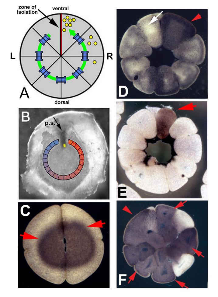
Gap junctions permit the direct passage of small molecules from the cytosol of one cell to that of its neighbor, and thus form a system of cell-cell communication that exists alongside familiar secretion/receptor signaling. Because of the rich potential for regulation of junctional conductance, and directional and molecular gating (specificity), gap junctional communication (GJC) plays a crucial role in many aspects of normal tissue physiology. However, the most exciting role for GJC is in the regulation of information flow that takes place during embryonic development, regeneration, and tumor progression. The molecular mechanisms by which GJC establishes local and long-range instructive morphogenetic cues are just beginning to be understood. This review summarizes the current knowledge of the involvement of GJC in the patterning of both vertebrate and invertebrate systems and discusses in detail several morphogenetic systems in which the properties of this signaling have been molecularly characterized. One model consistent with existing data in the fields of vertebrate left-right patterning and anterior-posterior polarity in flatworm regeneration postulates electrophoretically guided movement of small molecule morphogens through long-range GJC paths. The discovery of mechanisms controlling embryonic and regenerative GJC-mediated signaling, and identification of the downstream targets of GJC-permeable molecules, represent exciting next areas of research in this fascinating field.


Figures

Figure 1
Figure 1
Evidence for long-range GJC in chick and frog embryos determining left-right asymmetry Chick embryos cultured as whole blastoderms (A) exhibit the normal left-sided expression of the markers Nodal (B) and Sonic hedgehog in Hensen’s node (C), indicating normal asymmetry. In contrast, when the distal left side of the embryo is cut off before culture (D), cells on the far right side begin to express Nodal (E) and Sonic hedgehog expression in the node is bilateral (F). Panels A and D are schematics of st. 2 chick embryos (primitive streak stage); the region encompassed by the square indicates the part that was cultured. The purple stain in panels B-F is signal, indicating expression of the relevant gene marker via in situ hybridization. Red arrowheads indicate expression; white arrowheads indicate absence of expression. In frog embryos, small molecule fluorescent probes are able to penetrate several cell diameters through gap junctions (G). Panel G shows four large blastomeres of the 16-cell embryo. The left-most cell (indicated by red arrow) was injected with a fluorescent small molecule tracer dye, which is seen spreading to all 4 of its neighbors in a clock-wise direction. Panel H shows, in section, induced GJC (via injection of C×26 mRNA) in early frog embryos. The transfer of Lucifer Yellow (< 1kD) but not rhodamine-labeled dextran (RLD, =10 kD) in section demonstrates true gap junctional communication and rules out cytoplasmic bridges and incomplete cleavages.
Figure 2
Figure 2
Serotonin movement through gap junctions during LR patterning (A) One model consistent with the data is that in Xenopus, the long-range GJC path around the ventral zone of isolation results in a LR-asymmetric distribution of a small molecule morphogen (yellow). (B) The same model can be superimposed on the chick, where connexin43 is expressed circumferentially around, but not in, the primitive streak. (C) Serotonin is initially homogenously distributed in the early frog embryo. Within the next few hours, serotonin appears as a circumferential gradient around the zone (D) and ultimately coalesces into a single cell on one side of the midline (E). If the gap junctions are blocked, serotonin is unable to move (F). The frog embryo stages shown are 16-32-cell stages in panels A,D,E,F, and 2-cell stage in panel C. The chick embryo in panel B is at the early primitive-streak stage.
Figure 3
Figure 3
A model of electrophoretic movement of morphogens through gap-junctional paths (A) Schematically, a number of patterning systems can be visualized as a field of gap junctions terminating on a cell group at one end that establishes a strong polarization by ion exchange with the outside world. Gray level within the cells illustrate voltage gradient produced by pumps at the edge. (B) This results in an electrophoretic force that can, given realistic estimates of the physical parameters of cytoplasm and small molecules, result in a significant gradient of a small molecule across the cell field (yellow dots in panel A; this is now thought to be serotonin in the case of frog left-right patterning). (C) Importantly, mathematical analysis of the model illustrates that local gradients of GJC-permeable morphogens are thus set up within cells as well as across cell fields, potentially providing local directional cues.

References

    1. Aasen T, Hodgins MB, Edward M, Graham SV. The relationship between connexins, gap junctions, tissue architecture and tumour invasion, as studied in a novel in vitro model of HPV-16-associated cervical cancer progression. Oncogene. 2003;22:7969–80. - PubMed
    1. Adams DS, Robinson KR, Fukumoto T, Yuan S, Albertson RC, Yelick P, Kuo L, McSweeney M, Levin M. Early, H+-V-ATPase-dependent proton flux is necessary for consistent left-right patterning of non-mammalian vertebrates. Development. 2006;133:1657–1671. - PMC - PubMed
    1. Ai Z, Fischer A, Spray DC, Brown AM, Fishman GI. Wnt-1 regulation of connexin43 in cardiac myocytes. J Clin Invest. 2000;105:161–71. - PMC - PubMed
    1. Allen F, Tickle C, Warner A. The role of gap junctions in patterning of the chick limb bud. Development. 1990;108:623–34. - PubMed
    1. Araya R, Eckardt D, Maxeiner S, Kruger O, Theis M, Willecke K, Saez JC. Expression of connexins during differentiation and regeneration of skeletal muscle: functional relevance of connexin43. J Cell Sci. 2005;118:27–37. - PubMed

Publication types

LinkOut - more resources