. 2007 Jun 22;369(5):1258-69.
doi: 10.1016/j.jmb.2007.04.002. Epub 2007 Apr 5.

Structure of the FH2 domain of Daam1: implications for formin regulation of actin assembly

Affiliations

Structure of the FH2 domain of Daam1: implications for formin regulation of actin assembly

Jun Lu et al. J Mol Biol. .

Abstract

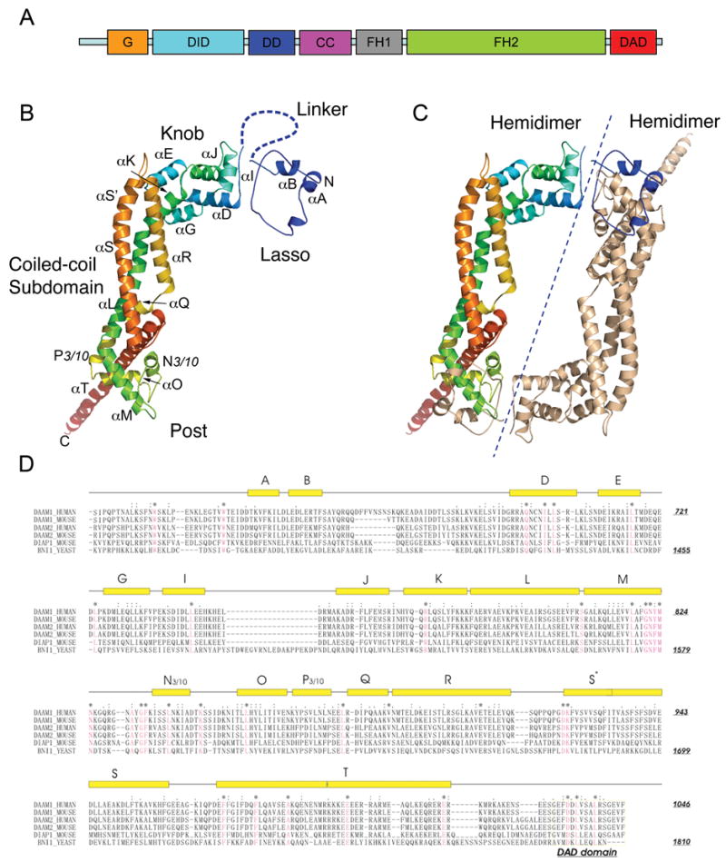
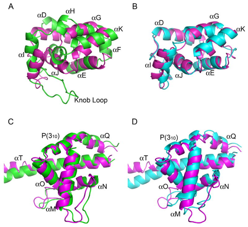
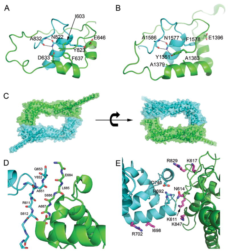
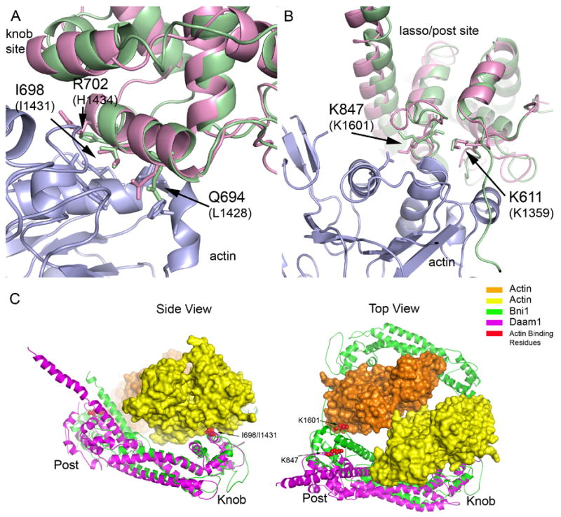
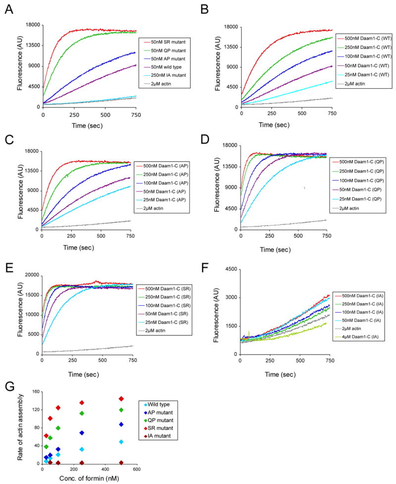
Daam1 (dishevelled-associated activator of morphogenesis-1) is a diaphanous-related formin first studied as a novel dishevelled binding protein and shown to be crucial for the planar cell polarity (PCP) pathway in Xenopus. Daam1, like other formins, directs nucleation and elongation of new actin filaments using its conserved formin-homology-2 (FH2) domain. Here we report the crystal structure of a large C-terminal fragment of human Daam1 containing the FH2 domain. The structure, determined at 2.25 A resolution using the single-wavelength anomalous diffraction (SAD) phasing method, reveals a "tethered dimer" architecture that is similar to that previously described for the FH2 domain of the yeast formin Bni1, which shares approximately 21% sequence identity with Daam1. Despite the overall similarity with the dimeric FH2 domain of Bni1 and with a truncated monomeric structure of mDia1, the Daam1 FH2 structure reveals a number of differences in secondary structure elements and in the "lasso/post" dimerization interface that may be functionally important. Most strikingly, the two halves of the crystallographic dimer pack together in a manner that occludes their actin binding surfaces. This "locked" conformation is stabilized by two novel, interacting beta-strands formed by the ends of the linkers that connect the two sides of the dimer. The Daam1 FH2 domain has weak actin assembly activity as compared with other mammalian formins, but mutations that disrupt the beta-strand lock increase activity about tenfold to a level comparable to other formins, suggesting that this occluded conformation may represent an auto-inhibited conformation of the Daam1 FH2 domain.


Figures

Figure 1
Figure 1
Crystal structure of the Daam1 FH2 domain. (A) The domain structure of human Daam1. (B) Ribbon diagram showing the overall structure of Daam1 FH2 domain. Subdomains including the lasso, the knob, the coiled-coil and the post region are labeled. The invisible linker region is drawn manually with dashed line for the purpose of illustration. The molecule is colored using the visible spectrum (from blue at the N-terminus to red at the C-terminus). (C) Ribbon diagram of the structure of Daam1 FH2 dimer. The dashed line separates the two hemidimers. One molecule is colored the same way as in (B), while the other is colored tan. (D) Sequence alignment and secondary structure of the FH2 and DAD domains. Aligned sequences are from human Daam1, murine Daam1, human Daam2, murine Daam2, murine Dia1 and yeast (S. cerevisiae) Bni1p. Secondary structure elements are shown above the sequences, with rectangles representing helices and thin lines indicating non-helical regions. Conserved residues are colored red. Figures wre prepared using the program PYMOL (Delano, W.L., The PyMol Molecular Graphics System (2002) http://www.pymol.org).
Figure 2
Figure 2
Structure comparisons of the FH2 domain of Daam1 with those of Bni1p and mDia1. (A–B) Comparisons of the knob subdomain of Daam1 and Bni1(A) or mDia1(B). (C–D) Comparisons of the post subdomain of Daam1 and Bni1(C) or mDia1(D). Daam1 is colored in magenta, Bni1 in green while mDia1 in cyan.
Figure 3
Figure 3
Distinct dimerization interactions in Daam1 and Bni1. Ribbon diagrams of (A) Daam1 and (B) Bni1 lasso/post interface. Conserved residues are labeled and shown in stick models. The lasso is colored in green and the post in cyan in (A–B). (C–E) The inter-hemidimer interface of Daam1 FH2 dimer. (C)Ribbon diagrams of Daam1 FH2 dimer are shown from the top view with one hemidimer colored in green and the other in cyan. The 50% transparent surfaces are also shown with the same color coding to emphasize the inter-hemidimer interface. (D–E) Ribbon diagrams of the inter-hemidimer interface. (D) A novel β-strand interaction formed by the two ends of the linker segment. (E) Residues expected to participate in actin binding are found at the inter-hemidimer interface. These residues are colored magenta and shown in stick form; they correspond to residues found at the actin binding site in the structure of the Bni1/actin complex. Mutation of the equivalent residues in Bni1 and/or mDia1 has been shown to diminish actin assembly activity. In particular, mutation of the residues corresponding to Lys847 and Ile698 completely abolish actin assembly activity in Bni1 (Lys1601 to Asp and Ile1431 to Ala) , . Note also that we show here that mutation of I698 to alanine abrogates the activity of the Daam1 FH2 domain (see Figure 5). Two residues from Daam1 (R692 and D740), which form a unique salt bridge, are also shown (cyan sidechains).
Figure 4
Figure 4
Insights into interactions with actin via comparisons with the structure of the Bni1/actin complex (A–B) The Daam1 FH2 structure is superimposed on the Bni1/actin complex (PDB ID 1Y64) in the regions of the knob actin-binding site (A) and the lasso/post binding site (B). The actin is colored blue, Daam1 magenta and Bni1 green. Selected residues that are known to be important for actin assembly by Bni1 are shown in stick form and labeled. (C) Overall views of the Daam1 FH2 domain (magenta) superimposed on the Bni1/actin complex (1Y64). Two actin subunits (yellow and orange) and two Bni1 FH2 domains (green) from the Bni1/actin structure are shown; this configuration may represent a “strained” intermediate in FH2-mediated assembly or actin filaments. The sidechains of key actin binding residues are shown in CPK form and are colored red (Ile698 and Lys847 in Daam1; Ile1431 and Lys1601 in Bni1). The superposition was carried out using the knob subdomain only. Note that while the knob and post sites independently superimpose well on the actin complex (panels A and B), both cannot be simultaneously brought into register with actin due to a different relative orientation of the knob subdomain. Bringing the knob into register (side view) leaves the actin binding residues in the laso/post region displaced from actin by ~17 Å (best seen in the Top view).
Figure 5
Figure 5
Actin assembly activities of wild type and mutant Daam1 FH2-C fragments. (A) Purified Daam1 polypeptides were directly compared at one concentration (50 nM) for their effects on assembly of 2 μM monomeric actin (5% pyrene-labeled). (B–F) Each purified Daam1 polypeptide was tested over a range of concentrations for its effects on assembly of 2 μM monomeric actin (5% pyrene-labeled). (G) Concentration-dependent effects of wild type and mutant Daam1 polypeptides on the rate of actin assembly. Rates were determined from the slopes of the actin assembly curves in B–F.

Similar articles

Cited by

References

    1. Kovar DR. Molecular details of formin-mediated actin assembly. Curr Opin Cell Biol. 2006;18:11–7. - PubMed
    1. Higgs HN. Formin proteins: a domain-based approach. Trends Biochem Sci. 2005;30:342–53. - PubMed
    1. Faix J, Grosse R. Staying in shape with formins. Dev Cell. 2006;10:693–706. - PubMed
    1. Goode BL, Eck MJ. Mechanism and Function of Formins in the Control of Actin Assembly. Annual Review of Biochemistry in press 2007 - PubMed
    1. Kohno H, Tanaka K, Mino A, Umikawa M, Imamura H, Fujiwara T, Fujita Y, Hotta K, Qadota H, Watanabe T, Ohya Y, Takai Y. Bni1p implicated in cytoskeletal control is a putative target of Rho1p small GTP binding protein in Saccharomyces cerevisiae. Embo J. 1996;15:6060–8. - PMC - PubMed

Publication types

MeSH terms

Associated data