. 2007 Jul;26(6):442-55.
doi: 10.1016/j.matbio.2007.03.002. Epub 2007 Mar 30.

The principal neuronal gD-type 3-O-sulfotransferases and their products in central and peripheral nervous system tissues

Affiliations

The principal neuronal gD-type 3-O-sulfotransferases and their products in central and peripheral nervous system tissues

Roger Lawrence et al. Matrix Biol. 2007 Jul.

Abstract

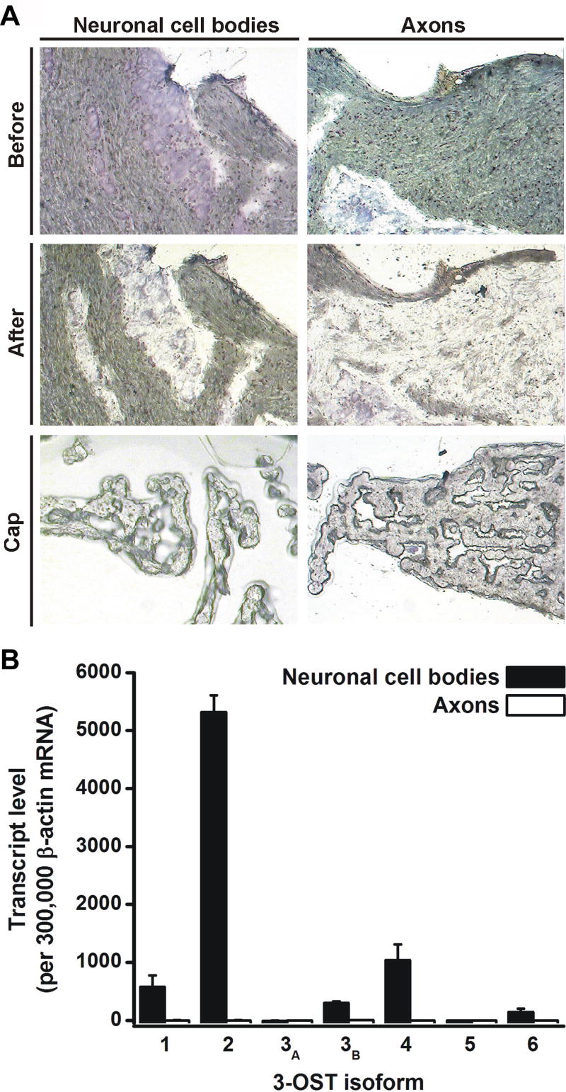
Within the nervous system, heparan sulfate (HS) of the cell surface and extracellular matrix influences developmental, physiologic and pathologic processes. HS is a functionally diverse polysaccharide that employs motifs of sulfate groups to selectively bind and modulate various effector proteins. Specific HS activities are modulated by 3-O-sulfated glucosamine residues, which are generated by a family of seven 3-O-sulfotransferases (3-OSTs). Most isoforms we herein designate as gD-type 3-OSTs because they generate HS(gD+), 3-O-sulfated motifs that bind the gD envelope protein of herpes simplex virus 1 (HSV-1) and thereby mediate viral cellular entry. Certain gD-type isoforms are anticipated to modulate neurobiologic events because a Drosophila gD-type 3-OST is essential for a conserved neurogenic signaling pathway regulated by Notch. Information about 3-OST isoforms expressed in the nervous system of mammals is incomplete. Here, we identify the 3-OST isoforms having properties compatible with their participation in neurobiologic events. We show that 3-OST-2 and 3-OST-4 are principal isoforms of brain. We find these are gD-type enzymes, as they produce products similar to a prototypical gD-type isoform, and they can modify HS to generate receptors for HSV-1 entry into cells. Therefore, 3-OST-2 and 3-OST-4 catalyze modifications similar or identical to those made by the Drosophila gD-type 3-OST that has a role in regulating Notch signaling. We also find that 3-OST-2 and 3-OST-4 are the predominant isoforms expressed in neurons of the trigeminal ganglion, and 3-OST-2/4-type 3-O-sulfated residues occur in this ganglion and in select brain regions. Thus, 3-OST-2 and 3-OST-4 are the major neural gD-type 3-OSTs, and so are prime candidates for participating in HS-dependent neurobiologic events.


Figures

Fig. 1
Fig. 1. Expression level of 3-OST isoforms in brain
Real-time RT-PCR of mouse brain total RNA was conducted using isoform specific primers for both first strand synthesis and PCR amplification. Transcript levels were calibrated against standard curves generated from cloned PCR products, and then standardized to the sample levels of β-actin, as described under “Experimental Procedures.” The results are expressed as the mean ± SEM for RNA samples extracted from eight separate mouse brains. 3-OST-6 expression levels, although low, were significantly above background reactions lacking reverse transcriptase.
Fig. 2
Fig. 2. Neuronal expression of 3-OST-2 and 3-OST-4 in the mouse trigeminal ganglia
In situ hybridizations with 3-OST-2 or 3-OST-4 antisense probes, as indicated, were performed on trigeminal ganglia longitudinal sections. Hybridization signals are most evident in the dark field images, seen as white grains. The corresponding hematoxylin/eosin stained bright field images show sensory ganglion cell bodies (upper portion of images) and an adjacent axon bundle (lower portion of images). Representative neurons (N), satellite cells (St), and Schwann cells (Sw) are indicated in these 400X images. For the 3-OST-2 probe, only scattered neurons exhibited significant hybridization signals (+). The remaining neurons (−) and non-neuronal cells exhibited a silver grain density that was comparable to control hybridizations with a sense probe (non shown). The 3-OST-4 signal is especially conspicuous over the majority of neuronal cell bodies. Reduced yet significant signal is also evident over satellite cells adjacent to highly expressive neurons (*). Silver grain density over axons was comparable to the background of the 3-OST-4 control sense probe (not shown)
Fig. 3
Fig. 3. 3-OST-2 and 3-OST-4 are the predominant 3-OST isoforms expressed by sensory neurons of the trigeminal ganglion
Individual sensory neurons with satellite cells (Neuronal cell bodies) or axonal processes with Schwann cells (Axons) were isolated from 8 μm cryosections of mouse trigeminal ganglia by LCM (Cap). Total RNA was extracted from samples containing approximately 100 neurons or 400 Schwann cells, then linearly-amplified RNA was subjected to real-time RT-PCR, as described under “Experimental Procedures”. (A) Hematoxylin and eosin stained sections (200X) before and after LCM, as indicated. For the neuronal samples, contaminating satellite cells comprised about 10% of the sample volume. (B) Expression of 3-OST isoforms in the indicated samples are the mean ± range from two independent cell isolations. Results are standardized to levels of β-actin.
Fig. 4
Fig. 4. Composite cDNA nucleotide and predicted amino acid sequences of human 3-OST-4
The cDNA sequence was compiled from overlapping genomic and cDNA sequences. Shown within the nucleic acid sequence are the polyadenylation signal (single underline) and a pure GC inverted repeat (double underline). Shown within the amino acid sequence are the transmembrane domain (boxed), the start of the sulfotransferase domain (arrow), and predicted sites for O-linked (*) and N-linked (dot underlined) glycosylation. Protein structural features were detected as previously described (Shworak et al., 1999). This sequence is GenBank accession number AF105378.
Fig. 5
Fig. 5. Identification of 3-OST-2/4-type products
HS, extracted from CHO[Eco] cells transduced with empty retroviral vector (Mock) or vectors expressing the indicated enzymes, was digested with Hep I-III and the derived residues were subjected to LC/MS analysis, as described under “Experimental Procedures”. Enzymatic cleavage converts GlcA/IdoA to ΔUA. For simplicity, the data are presented as the extracted ion current for sulfated HS saccharide residues only. Residues common to all chromatograms are indicated in the top (Mock) panel. 3-O-sulfated residues resulting from retroviral transduction are indicated in the 3-OST-2 and 3-OST-4 panels. Retention times varied slightly between duplicate runs of the same sample, so peak assignments were confirmed by their mass spectrographs (not shown), as we have previously described (Lawrence et al., 2004). Shown to the right are enlargements of the 62 – 70 min region with separate tracings indicating compounds having m/z values (± 0.2 atomic mass units) consistent with sulfated tetrasaccharide residues (solid line) or the fully sulfated disaccharide, ΔUA2S→GlcNS3S6S (broken line). The open arrow indicates apparent contaminating species having m/z values inconsistent with but within the tolerance set for HS saccharide residues.
Fig. 6
Fig. 6. 3-OST-2/4-type HS products occur in select neural tissues
LC/MS analysis of Hep I-III digested HS extracted from the indicated mouse tissues. For simplicity we present data for two diagnostic molecular ions. The chromatograms show the extracted ion current for compounds having m/z values (± 0.2 atomic mass units) consistent with ΔUA2S→GlcNS3S6S and Tetra B. Beneath are mass spectra for the [M-2H+DBA]-1 form of ΔUA2S→GlcNS3S6S and the [M-2H]-2 form of Tetra B. For the Tetra-B [M-2H]-2 molecular ion (m/z of 536 atomic mass units), the -2 charge is confirmed by the isotopic cluster, which shows 0.5 atomic mass unit difference between adjacent peaks. For the ΔUA2S→GlcNS3S6S [M-2H+DBA]-1 molecular ion (m/z of 785 atomic mass units), the isotopic cluster progresses by 1 atomic mass unit, indicating the charge of -1. The trigeminal sample also contains a contaminant (masses indicated in italics) co-eluting within the ΔUA2S→GlcNS3S6S region, which is distinguished by its m/z of 786.6 atomic mass units and its charge of -2 (peaks differing by 0.5 atomic mass units). The 787.1 peak likely contains signal from both the 3-O-sulfated disaccharide and the contaminant. For the cerebellum, the mass spectra corresponding to the elution ranges seen in both the trigeminal and the brain stem verified the absence of these species in cerebellar HS.
Fig. 7
Fig. 7. 3-OST-2 and 3-OST-4 expression converts resistant CHO cells to susceptibility to HSV-1 entry
CHO cells were transfected with plasmids expressing the indicated 3-OST constructs or the empty vector (pcDNA3.1). Transfectants were exposed to serial dilutions of HSV-1(KOS)tk12, which expresses β-galactosidase from an insert in the viral genome. Viral entry-dependent β-galactosidase activity was quantitated at 6 h after the addition of virus, as described under “Experimental Procedures”. Representative values from a single transfection show the amount of reaction product detected spectrophotometrically (A405) as the mean from 3 wells per virus dose. Standard deviations were typically <15% of the mean. Comparable results were obtained in 5 independent transfection experiments.
Fig. 8
Fig. 8. The 3-OST multigene family is comprised of two structurally and functionally defined groups
The dendrogram of the human 3-OST multigene family shows the degree of sequence homology (% Similarity) within the previously defined sulfotransferase domain (Shworak et al., 1999; Yabe et al., 2001). Indicated are the preferred HS motifs generated by each enzyme, gD- or antithrombin (AT)-binding sites, (Shukla et al., 1999; Shworak et al., 1996; Shworak et al., 1997; Xia et al., 2002; Xu et al., 2005, and data herein), and the resulting structural/functional categories. The dendrogram was generated with the University of Wisconsin Genetics Computer Group program “Distances” using amino acid sequences from GenBank/EMBL/DDBJ accession numbers: AF019386 (3-OST-1), AF105374 (3-OST-2), AF105376 (3-OST-3A), AF105377 (3-OST-3B), AF105378 (3-OST-4), AF50392 (3-OST-5), and AY574375 (3-OST-6).

References

    1. Alvarez-Buylla A, Lim DA. For the long run: maintaining germinal niches in the adult brain. Neuron. 2004;41:683–686. - PubMed
    1. Artavanis-Tsakonas S, Rand MD, Lake RJ. Notch signaling: cell fate control and signal integration in development. Science. 1999;284:770–776. - PubMed
    1. Borjigin J, Deng J, Sun X, De Jesus M, Liu T, Wang MM. Diurnal pineal 3-O-sulphotransferase 2 expression controlled by beta-adrenergic repression. J Biol Chem. 2003;278:16315–16319. - PubMed
    1. Bulow HE, Berry KL, Topper LH, Peles E, Hobert O. Heparan sulfate proteoglycan-dependent induction of axon branching and axon misrouting by the Kallmann syndrome gene kal-1. Proc Natl Acad Sci U S A. 2002;99:6346–6351. - PMC - PubMed
    1. Chatterton JE, Hirsch D, Schwartz JJ, Bickel PE, Rosenberg RD, Lodish HF, Krieger M. Expression cloning of LDLB, a gene essential for normal Golgi function and assembly of the ldlCp complex. Proc Natl Acad Sci U S A. 1999;96:915–920. - PMC - PubMed

Publication types

MeSH terms

Associated data

LinkOut - more resources