Review
. 2007 Nov;64(2):159-71.
doi: 10.1016/j.critrevonc.2007.03.001. Epub 2007 May 4.

Role of erythropoietin in the brain

Affiliations
Review

Role of erythropoietin in the brain

Constance Tom Noguchi et al. Crit Rev Oncol Hematol. 2007 Nov.

Abstract

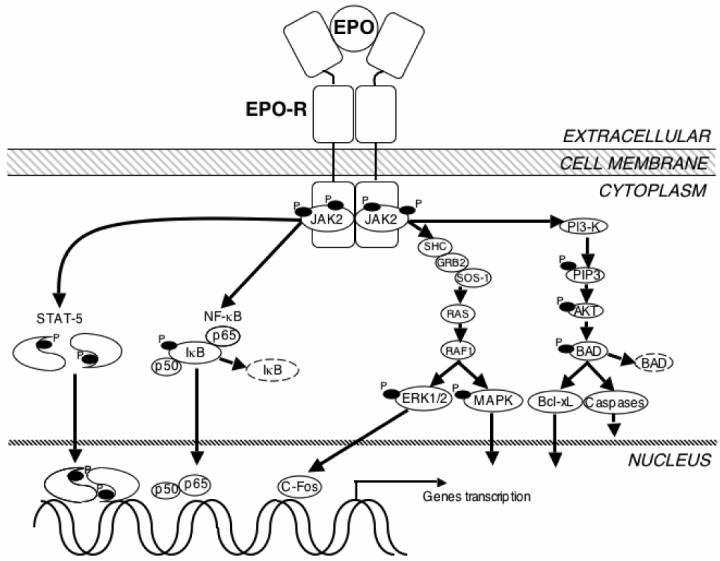
Multi-tissue erythropoietin receptor (EPO-R) expression provides for erythropoietin (EPO) activity beyond its known regulation of red blood cell production. This review highlights the role of EPO and EPO-R in brain development and neuroprotection. EPO-R brain expression includes neural progenitor cells (NPC), neurons, glial cells and endothelial cells. EPO is produced in brain in a hypoxia sensitive manner, stimulates NPC proliferation and differentiation, and neuron survival, and contributes to ischemic preconditioning. Mice lacking EPO or EPO-R exhibit increased neural cell apoptosis during development before embryonic death due to severe anemia. EPO administration provides neural protection in animal models of brain ischemia and trauma, reducing the extent of injury and damage. Intrinsic EPO production in brain and EPO stimulation of endothelial cells contribute to neuroprotection and these are of particular importance since only low levels of EPO appear to cross the blood-brain barrier when administered at high dose intravenously. The therapeutic potential of EPO for brain ischemia/trauma and neurodegenerative diseases has shown promise in early clinical trial and awaits further validation.


Figures

Figure 1
Figure 1
EPO signaling in neural cells. In neural cells, EPO binds to the EPO-R dimmer and stimulates JAK2 kinase activity resulting in phosphorylation (P) of JAK2 and EPO-R. Activated JAK2 initiates signal transduction through several adaptor proteins such as Src homology containing protein (SHC), growth factor receptor-binding protein 2 (GRB2), son of sevenless protein-1 (SOS-1), and phosphoinositol 3-kinase (PI3-K). The downstream signaling messengers include the G protein (RAS), serine/threonine-specific kinase (RAF1), p38 mitogen-activated protein kinase (MAPK), extracellular response– stimulated kinase (ERK1/2), human oncogene (C-Fos), membrane phospholipids (phosphoinositol 3,4,5-phosphate, PIP3), protein kinase B (AKT), apoptosis inducer (BAD), anti-apoptotic protein (Bcl-xL), and the caspases. Additional signaling pathways contain signal transducer and activator of transcription 5 (STAT5) and Nuclear factor kappa B (NF-κB). All of these pathways have been found to affect the gene transcription in neural cells survive related to EPO.
Figure 2
Figure 2
Contributions of EPO activity to neuroprotection and repair. Erythroid progenitor cells, endothelial cells, neural progenitor cells and neurons express EPO-R. Direct EPO stimulation of neural progenitor cells or neurons can be regenerative or neuroprotective. Indirectly, EPO can increase oxygen delivery to the brain through stimulation of erythroid progenitor cells to increase mature red blood cell production and stimulation of endothelial cells to increase NO production. EPO stimulated endothelial cells can indirectly promote neural cell survival via production of neurotrophic factors.
Figure 3
Figure 3
EPO neuroprotection in ischemic brain injury. Neural progenitor cells and neurons can receive EPO in three ways: 1) Increased production of EPO by autocrine action or from adjacent neurons, 2) Increased production of EPO by glial cells, e.g. astrocytes, 3) Increased EPO crossing the blood- brain barrier in which the permeability is comprised during hypoxia/ischemia. In addition, EPO may stimulate endothelial cells to secrete neurotrophic factors and hence provide indirect neuroprotection.

References

    1. Anagnostou A, Liu Z, Steiner M, et al. Erythropoietin receptor mRNA expression in human endothelial cells. Proc Natl Acad Sci U S A. 1994;91(9):3974–8. - PMC - PubMed
    1. Anagnostou A, Lee ES, Kessimian N, Levinson R, Steiner M. Erythropoietin has a mitogenic and positive chemotactic effect on endothelial cells. Proc Natl Acad Sci U S A. 1990;87(15):5978–82. - PMC - PubMed
    1. Morishita E, Masuda S, Nagao M, Yasuda Y, Sasaki R. Erythropoietin receptor is expressed in rat hippocampal and cerebral cortical neurons, and erythropoietin prevents in vitro glutamate-induced neuronal death. Neuroscience. 1997;76(1):105–16. - PubMed
    1. Jelkmann W, Wagner K. Beneficial and ominous aspects of the pleiotropic action of erythropoietin. Ann Hematol. 2004;83(11):673–86. - PubMed
    1. Wu H, Liu X, Jaenisch R, Lodish HF. Generation of committed erythroid BFU-E and CFU-E progenitors does not require erythropoietin or the erythropoietin receptor. Cell. 1995;83(1):59–67. - PubMed