. 2007 May 4;129(3):579-91.
doi: 10.1016/j.cell.2007.03.023.

Spinophilin facilitates dephosphorylation of doublecortin by PP1 to mediate microtubule bundling at the axonal wrist

Affiliations

Spinophilin facilitates dephosphorylation of doublecortin by PP1 to mediate microtubule bundling at the axonal wrist

Stephanie L Bielas et al. Cell. .

Erratum in

  • Cell. 2007 Jun 15;129(6):1227-8

Abstract

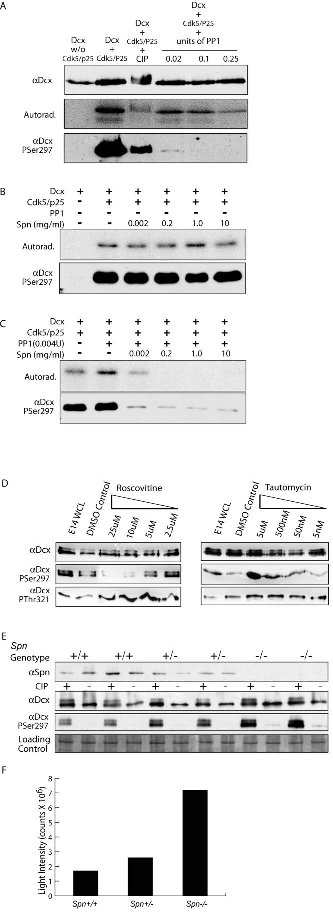
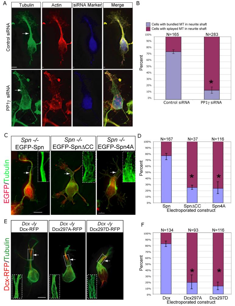
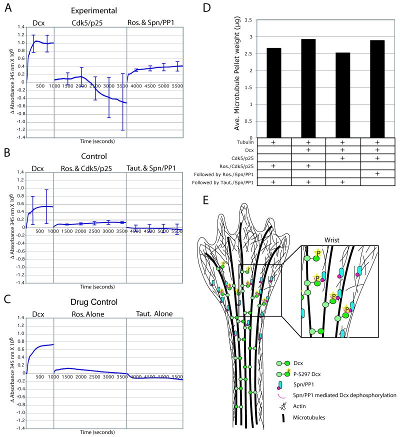
The axonal shafts of neurons contain bundled microtubules, whereas extending growth cones contain unbundled microtubule filaments, suggesting that localized activation of microtubule-associated proteins (MAP) at the transition zone may bundle these filaments during axonal growth. Dephosphorylation is thought to lead to MAP activation, but specific molecular pathways have remained elusive. We find that Spinophilin, a Protein-phosphatase 1 (PP1) targeting protein, is responsible for the dephosphorylation of the MAP Doublecortin (Dcx) Ser 297 selectively at the "wrist" of growing axons, leading to activation. Loss of activity at the "wrist" is evident as an impaired microtubule cytoskeleton along the shaft. These findings suggest that spatially restricted adaptor-specific MAP reactivation through dephosphorylation is important in organization of the neuronal cytoskeleton.


Figures

Figure 1
Figure 1
Delayed axonal extension in Dcx −/y brains. (A) Midline sagittal T1-weighted brain MRI from normal showed well-formed CC and a male with deletion of DCX exon 7-8 showed severe CC hypoplasia (arrows). Optic nerve (arrowhead), cerebellum (*), cortex (**). (B) E14.5 DiI injected into medial subcortical region showed extensive fibers in subcortical white matter (arrow), cortico-striatal (CS) boundary (double arrows) and striatal-thalamus region (triple arrows). Mutant showed minimal axonal extension from the injection. (C) E15.5 DiI injection showed labeling of corticothalamic (CT) axons at CS boundary (box 1) and striatal-thalamus region (box 2), whereas mutant showed diminished labeling. (D) High power views. (E) E16.5 DiI injection showed diminished axon extension of the CC tract (arrowhead) in mutant. There was some catch-up extension of CT tract by this age (arrow).
Figure 2
Figure 2
Spn and Dcx share protein distribution and co-function during brain development. (A) Dcx exhibited enrichment along MTs at the wrist (double arrows), the axonal shaft (arrowhead) and cell body (double arrowheads), PP1 was distributed diffusely, and Spn was enriched at the wrist (double arrows). PSer297 Dcx had highest expression in the growth cone (arrow) and cell body (double arrowhead). PSer297 Dcx compared with total Dcx was mostly excluded from regions of Spn localization at the wrist (double arrow), suggesting Spn may contribute to its dephosphorylation. Scale bar 10 μm. (B) Dcx and Spn overlap in distribution with the actin and MT cytoskeletons at the wrist (double arrow) and filopodia (arrowheads). (C) Dcx and Spn cooperate in axonal outgrowth of multiple long distance projections. Both Spn −/− and Dcx −/y showed defective lamination of the CA3 region (arrow), whereas Spn −/−; Dcx −/y (DKO) showed possibly worsened defect compared with single knockouts. The granule cell layer was unaffected (arrowhead). The CC decussation was evident in all but the DKO, where it was replaced by Probst bundles (PB). The anterior commissure (dashes) showed normal appearance in all but the DKO where it was hypoplastic. Midline indicated by arrowhead. At P0, decussating CC fibers (stained with L1CAM, arrows) were visible in all but the DKO, where they terminated in Probst bundles (arrowheads). (D) Expressivity of ACC and hypoplastic anterior commissure among offspring from 20 litters of double heterozygous matings. Number of mice with each phenotype over total of each genotype are listed. Note that none of the mice except the Spn −/− ; Dcx −/− (DKO) showed ACC and hypoplastic anterior commissure phenotype. Dcx −/− entries include both −/− females and −/y male null mice. * = p < 0.001, Chi squared test.
Figure 3
Figure 3
Disrupted shaft MT cytoskeleton in Dcx, Spn and DKO mutant neurons results in excessively branched neurite phenotype. (A) WT showed condensed MTs in the shaft (arrowhead). Wrist (double arrow) and growth cone (arrow) are well-delineated from the cell body (dashed circle = nucleus). In Dcx −/y or Spn/− neurons, MTs were instead splayed (arrowhead), and as a result, the neurite shaft width was increased. (B) Transmission electron microscopy showed parallel MT arrays that maintained relatively consistent spacing along the shafts in cultured cortical neurons in wt littermates. This array was disrupted in both single knockouts and was more severe in the DKO. Bottom half (dashes) of each shows line tracings depicting MTs. 8000X. (C) Binned inter-MT distance between nearest neighbor was significantly greater in mutants. N = 1499 total measurements, from 9 wt, 5 Dcx−/−, 4 Spn −/− and 5 DKO neuronal shafts from two separate culture experiments. Error bar = SEM. * = p < 0.05, Chi squared contingency table. (D) WT neurons typically displayed a single monopolar main process (arrow) with occasional 2° branches (double arrow) after 36 hrs in culture. Both Dcx −/y and Spn −/y neurons exhibited excessive 2° (double arrows) and 3° (triple arrows) branches from the primary neurite, as well as an increased number of processes extending from the cell soma (arrowhead). This was most striking in DKO. (E) Quantification of neurite branching. Branches from the main process (MP) were termed 2° MP, and branches from 2° MP were termed 3° MP. Neurites extending from the soma were termed body processes (BP), and branches from the BP were termed 2° BP. (F) WT cells typically had a single MP, with average number of 2° MP per cell less than 0.5. The branching and frequency of 3° MP was increased in Dcx −/y, Spn −/− and DKO neurons. Furthermore, 2° BP were only noted in DKO neurons. * = p < 0.05, pairwise comparison, Student t-test.
Figure 4
Figure 4
Dcx/Spn interaction sufficient to co-recruit actin and MT cytoskeletons. Purified Spn and Dcx was sufficient to link phalloidin-stabilized actin and taxol-stabilized MTs. (A-B) Spn added to actin led to production of filaments. (C-D) Dcx added to tubulin led to asters of MTs. (E) Cytoskeletons alone showed no co-recruitment, and neither was tubulin recruited to Spn-stabilized actin (F) or actin recruited to Dcx-stabilized tubulin (G). However, Spn-stabilized actin and Dcx-stabilized MTs showed significant co-recruitment of the two cytoskeletons (H, and higher power view of H). Repeated in triplicate.
Figure 5
Figure 5
Spn required for PP1-mediated dephosphorylation of PSer297 Dcx. (A) PP1 at high unit concentrations was capable of dephosphorylating Dcx at PSer297, based on autoradiogram or immunoreactivity with αPSer297 following [32P] incorporation. PP1 was a more specific phosphatase for the PSer297 site than CIP, resulting in nearly complete dephosphorylation at all concentrations tested. (B) Spn alone has no effect on [32P] retention or αPSer297 reactivity. (C) Low levels of PP1 (5X lower than used in (A) in the absence of Spn had no effect on [32P] retention or αPSer297 reactivity but increasing amounts of Spn promoted dephosphorylation of Dcx by PP1. (D) PP1 and Cdk5 act in opposing fashions to modulate phosphorylation state of Dcx Ser297 in cortical neurons. Cortical neurons with increasing roscovitine (inhibits Cdk5) or tautomycin (inhibits PP1) were analyzed by Western with Dcx PSer297 and PThr321 antibodies. Roscovitine blocked and tautomycin enhanced PSer297 reactivity but not PThr321. (E) Brain lysates from E16 littermates showed increased PSer297 reactivity as Spn dosage was decreased. (F) Quantification of PSer297 Dcx band intensity standardized to control shows a four-fold increase in reactivity in Spn −/− versus +/+.
Figure 6
Figure 6
Spn-PP1-Dcx complex required for MT bundling during neurite outgrowth. (A) PP1γ knockdown associated with failure of MT bundling and broadened primary neurite. (C) Spn −/− splayed MT phenotype is rescued by forced expression of EGFP-tagged wildtype Spn, but not SpnΔCC (lacks Dcx binding) or Spn4A (lacks PP1 binding). (E) Dcx −/y splayed MT phenotype is rescued by forced expression of Dcx-RFP, but not by Dcx297A (unphosphorylatable) or Dcx297D (pseudophosphorylated) at the Spn-PP1 site. Scale bar 5 μm. (B, D, F) Significant difference in cells with bundled vs. splayed MT neurite phenotype. Results averaged from two experiments. * = p < 0.01, Student t-test.
Figure 7
Figure 7
Spn-PP1-mediated dephosphorylation reinstates the tubulin polymerization effect of Dcx. (A) Purified Dcx and tubulin shows robust increase in turbidity after 1000 seconds. Subsequently, addition of activated Cdk5/p25 to phosphorylate Ser297 resulted in a net decrease in turbidity over the next 2500 sec. Subsequently, addition of roscovitine (to block Cdk5 activity) and Spn-PP1 (to dephosphorylate PSer297 Dcx) resulted in net increase in turbidity over the next 2500 sec. (B) Cdk5/p25 that was pretreated with roscovitine, or Spn-PP1 that was pretreated with tautomycin had no net effect on turbidity. (C) Neither roscovitine nor tautomycin alone had any net effect on turbidity. Error bars = SEM from three trials. (D) Co-sedimentation analysis. The dephosphorylation of previously phosphorylated Dcx sites was associated with a reinstatement of Dcx MT polymerizing activity, and associated with an increased MT pellet weight. Averaged from two experiments. (E) Model for the role of Dcx and Spn in MT organization during neurite extension. Spn is restricted to the wrist region, where it is complexed with PP1. Spn mediates PP1 dephosphorylation of MT-bound PSer 297 Dcx. This leads to reactivation of Dcx, with subsequent MT crosslinking activity that is necessary for MT bundling in the neurite shaft.

References

    1. Agarwal-Mawal A, Paudel HK. Neuronal Cdc2-like protein kinase (Cdk5/p25) is associated with protein phosphatase 1 and phosphorylates inhibitor-2. J Biol Chem. 2001;276:23712–23718. - PubMed
    1. Allen PB, Ouimet CC, Greengard P. Spinophilin, a novel protein phosphatase 1 binding protein localized to dendritic spines. Proc Natl Acad Sci U S A. 1997;94:9956–9961. - PMC - PubMed
    1. Arlotta P, Molyneaux BJ, Chen J, Inoue J, Kominami R, Macklis JD. Neuronal subtype-specific genes that control corticospinal motor neuron development in vivo. Neuron. 2005;45:207–221. - PubMed
    1. Ceulemans H, Bollen M. Functional diversity of protein phosphatase-1, a cellular economizer and reset button. Physiol Rev. 2004;84:1–39. - PubMed
    1. Corbo JC, Deuel TA, Long JM, LaPorte P, Tsai E, Wynshaw-Boris A, Walsh CA. Doublecortin is required in mice for lamination of the hippocampus but not the neocortex. J Neurosci. 2002;22:7548–7557. - PMC - PubMed

Publication types

MeSH terms