. 2007 Sep;37(11):1221-32.
doi: 10.1016/j.ijpara.2007.03.005. Epub 2007 Mar 30.

Minimum requirements for ookinete to oocyst transformation in Plasmodium

Affiliations

Minimum requirements for ookinete to oocyst transformation in Plasmodium

Victoria Carter et al. Int J Parasitol. 2007 Sep.

Abstract

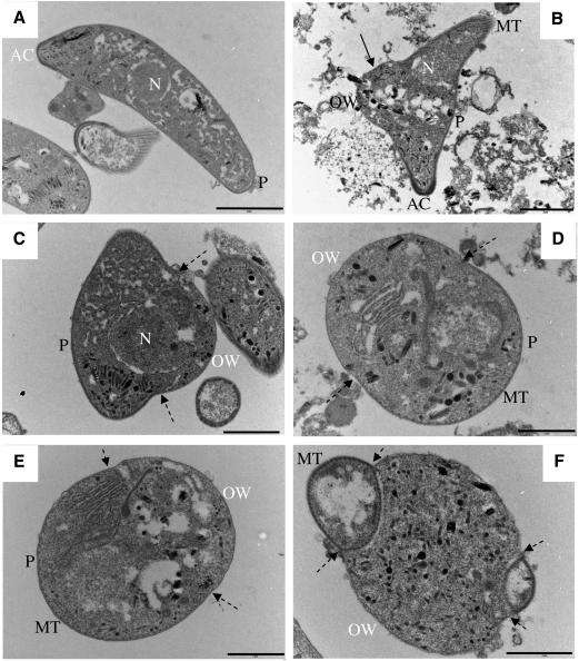
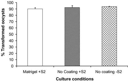
During their passage through a mosquito vector, malaria parasites undergo several developmental transformations including that from a motile zygote, the ookinete, to a sessile oocyst that develops beneath the basal lamina of the midgut epithelium. This transformation process is poorly understood and the oocyst is the least studied of all the stages in the malaria life cycle. We have used an in vitro culture system to monitor morphological features associated with transformation of Plasmodium berghei ookinetes and the role of basal lamina components in this process. We also describe the minimal requirements for transformation and early oocyst development. A defined sequence of events begins with the break-up of the inner surface membrane, specifically along the convex side of the ookinete, where a protrusion occurs. A distinct form, the transforming ookinete or took, has been identified in vitro and also observed in vivo. Contrary to previous suggestions, we have shown that no basal lamina components are required to trigger ookinete to oocyst transformation in vitro. We have demonstrated that transformation does not occur spontaneously; it is initiated in the presence of bicarbonate added to PBS, but it is not mediated by changes in pH alone. Transformation is a two-step process that is not completed unless a range of nutrients are also present. A minimal medium is defined which supports transformation and oocyst growth from 7.8 to 11.4microm by day 5 with 84% viability. We conclude that ookinete transformation is mediated by bicarbonate and occurs in a similar manner to the differentiation of sporozoite to the hepatic stage.


Figures

Fig. 1
Fig. 1
Formation of young Plasmodium berghei oocysts in vitro. (A–C) P28 (surface) and DAPI (nuclear) staining of ookinetes (A), tooks (B) and young oocysts (C). (D) PbGFPCON tooks during late transformation in culture. (E) PbGFPCON early transforming ookinetes in Anopheles stephensi midguts 27 h post-blood feeding (composite of three images). (F) Image adapted from Sinden et al. (1987) (i–iv) to incorporate ookinete to oocyst transformation (v–vii). Transformation begins with a small hump forming on the outer, convex, edge of the ookinete, often towards the apical end. Transforming ookinetes (tooks) then take on a snail-like appearance (v) and the nucleus is observed to move into the developing oocyst. Remnants of the ookinete tips are clearly visible (distinguishing these forms from zygotes; (vi) just prior to the formation of spherical young oocysts. The entire population of ookinetes transform in 12–36 h, depending on nutrient availability.
Fig. 2
Fig. 2
TEM images of an ookinete (A), and tooks (B–F) in vitro. (A) Ookinete, with nucleus (N), pellicle (P) and apical complex (AC) visible. (B) Early took: the AC is still clearly visible as are the microtubules (MT) and the beginning of the early oocyst wall, forming a hump on the outer ookinete edge. Membrane generated during this process differs from the ookinete pellicle (single arrow), and forms the basis of the oocyst wall (OW). (C) Early took: nuclear material can be seen to have moved into the newly formed area. Double and single membranes define the new OW and ookinete pellicle. Broken arrows indicate the transition areas from double to newly formed single membranes. (D and E) Mid-stage tooks showing microtubules below the remaining pellicle. (F) Late took: remnants of the ookinete tips with double membranes can be seen at either end of the forming oocyst: scale bar: A–C, 2 μm; D–F, 1 μm.
Fig. 3
Fig. 3
Production of laminin by Drosophila melanogaster S2 cells. Cells were cultured at 27 °C in Schneider’s Insect Cell Medium supplemented with FBS and Pen/Strep. (A) Western blot to visualise laminin secreted into the medium. The supernatant from a confluent flask was subjected to SDS–PAGE and transferred to a nitrocellulose membrane. The blots were probed with anti-D. melanogaster laminin-1 (β- and γ-chains) polyclonal antibodies (lane 1). Molecular markers are shown on the right (lane 2) and the corresponding molecular weight labelled. (B) Laminin secretion by S2 cells was determined by ELISA and readings are expressed as cell equivalents per 100 μl of medium. Error bars = SEM and n = 3 wells.
Fig. 4
Fig. 4
Maximal ookinete to oocyst transformation rates 24-h post-culture. Transformation rates were assessed by counting the number of parasites out of 100 that had changed from ookinete shape to young oocysts. No significant difference (ANOVA, F(2,2) = 0.90, P = 0.422) was observed between full-culture conditions, culture without Matrigel and culture without Matrigel and Drosophila S2 cells. Bars represent SEM. The experiment was repeated three times and parasites counted in three separate wells for each condition.

References

    1. Adini A., Warburg A. Interaction of Plasmodium gallinaceum ookinetes and oocysts with extracellular matrix proteins. Parasitology. 1999;1999:331–336. - PubMed
    1. Al-Olayan E.M., Beetsma A.L., Butcher G.A., Sinden R.E., Hurd H. Complete development of mosquito phases of the malaria parasite in vitro. Science. 2002;295:677–679. - PubMed
    1. Al-Olayan E.M., Williams G.T., Hurd H. Apoptosis in the malaria protozoan, Plasmodium berghei: a possible mechanism for limiting intensity of infection in the mosquito. Int. J. Parasitol. 2002;32:1133–1143. - PubMed
    1. Arai M., Billker O., Morris H.R., Panico M., Delcroix M., Dixon D., Ley S.V., Sinden R.E. Both mosquito-derived xanthurenic acid and a host blood-derived factor regulate gametogenesis of Plasmodium in the midgut of the mosquito. Mol. Biochem. Parasitol. 2001;116:17–24. - PubMed
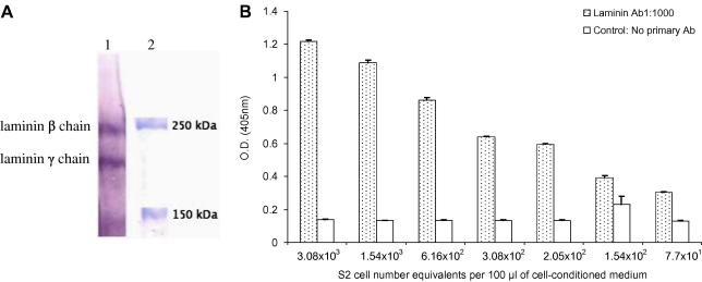
    1. Arrighi R.B., Lycett G., Mahairaki V., Siden-Kiamos I., Louis C. Laminin and the malaria parasite’s journey through the mosquito midgut. J. Exp. Biol. 2005;208:2497–2502. - PubMed

Publication types

LinkOut - more resources