. 2007 Jul;91(3):239-50.
doi: 10.1016/j.ymgme.2007.03.001. Epub 2007 May 7.

Bone marrow transplantation for feline mucopolysaccharidosis I

Affiliations

Bone marrow transplantation for feline mucopolysaccharidosis I

N Matthew Ellinwood et al. Mol Genet Metab. 2007 Jul.

Abstract

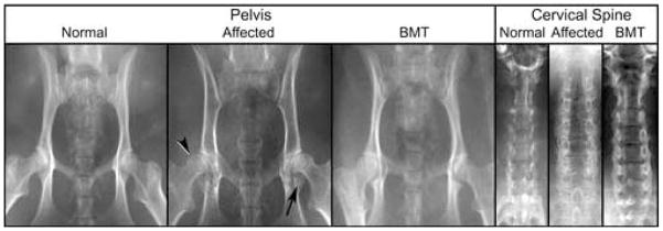
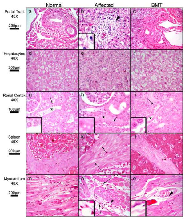
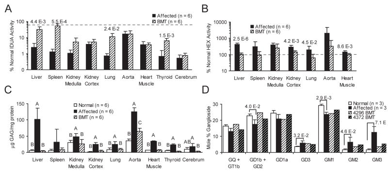
Severe mucopolysaccharidosis type I (MPS I) is a fatal neuropathic lysosomal storage disorder with significant skeletal involvement. Treatment involves bone marrow transplantation (BMT), and although effective, is suboptimal, due to treatment sequelae and residual disease. Improved approaches will need to be tested in animal models and compared to BMT. Herein we report on bone marrow transplantation to treat feline mucopolysaccharidosis I (MPS I). Five MPS I stably engrafted kittens, transplanted with unfractionated bone marrow (6.3x10(7)-1.1x10(9) nucleated bone marrow cells per kilogram) were monitored for 13-37 months post-engraftment. The tissue total glycosaminoglycan (GAG) content was reduced to normal levels in liver, spleen, kidney, heart muscle, lung, and thyroid. Aorta GAG content was between normal and affected levels. Treated cats had a significant decrease in the brain GAG levels relative to untreated MPS I cats and a paradoxical decrease relative to normal cats. The alpha-l-iduronidase (IDUA) activity in the livers and spleens of transplanted MPS I cats approached heterozygote levels. In kidney cortex, aorta, heart muscle, and cerebrum, there were decreases in GAG without significant increases in detectable IDUA activity. Treated animals had improved mobility and decreased radiographic signs of disease. However, significant pathology remained, especially in the cervical spine. Corneal clouding appeared improved in some animals. Immunohistochemical and biochemical analysis documented decreased central nervous system ganglioside storage. This large animal MPS I study will serve as a benchmark of future therapies designed to improve on BMT.


Figures

Figure 1
Figure 1. Composite Radiograph of MPS I Normal, Affected, and BMT-treated cats
Images of the pelvis show characteristic changes in MPS I affected cats: coxofemoral joint subluxation (arrowhead), and acetabular flattening with bony proliferation (arrow). These changes are reduced in the BMT treated animal. The untreated MPS I cervical spine shows increased vertebral width and bony proliferation, both of which are reduced in the BMT treated animal.
Figure 2
Figure 2. Histopathology of Somatic Tissues
Hematoxylin and eosin-stained sections from normal, MPS I, and MPS I BMT-treated cats. Tissue, magnification, and micron bars located to the left of designated rows. Panels a, b, and c (liver portal tract): arrowhead (b) indicates vacuolated mononuclear cell infiltration of portal tract (3X inset) in an untreated MPS I cat, a finding absent in normal and BMT-treated cats. Panels d, e, and f (hepatocytes): the affected cat has enlarged hepatocytes with many small uniform cytoplasmic vacuoles. Normal and BMT samples have vacuolation typical of normal feline liver, are similar to each other, and differ strikingly from affected. Panels g, h, and i (renal cortex): asterisks indicate distal convoluted tubules. Cytoplasmic vacuoles in affected distal convoluted tubules are absent in normal and BMT cats (arrows indicate 3X insets). Panels j, k, and l (splenic trabeculae). Vacuolation of smooth muscle cells in MPS I (arrows) is decreased in BMT-treated cats. Panels m, n, and o (myocardium): arrows indicate perivascular infiltration by mononuclear cells in an affected cat, which is absent in the BMT animal. Arrowheads indicate 3X insets in panels n and o.
Figure 3
Figure 3. Histopathology of CNS Tissues
Hematoxylin and eosin-stained sections from normal, MPS I, and MPS I BMT-treated cats. Tissue, magnification, and micron bars located to the left of designated rows. Panels a, b, and c (cortical neurons): affected and BMT cortical neurons are swollen with cytoplasmic vacuoles, however BMT neurons were slightly less so (insets are 2X images of neurons indicated by arrows). Panels d, e, and f (hippocampal neurons): severity of neuronal cytoplasmic vacuolation is decreased in BMT (insets are 4X images of neurons indicated by arrows). Panels g, h, and I (cerebral perivascular space): number of vacuolated mononuclear cells (arrow, panel h) is nearly absent in BMT (arrow, panel i). Panels j, k, and l (choroid plexus): infiltration of choroid plexus (asterisks) by vacuolated mononuclear cells and cytoplasmic vacuolation of epithelial cuboidal cells (arrows indicate 4X insets) present in affected cat are nearly absent in BMT-treated cat. Panels m, n, and o (leptomeninges): vacuolation of fibroblasts and mononuclear cell infiltrates in affected cat are nearly absent in BMT-treated cat (arrows).
Figure 4
Figure 4. GM2and GM3Ganglioside Storage in Supragranular Cerebral Cortex
ICC stained sections from normal, MPS I, and MPS I BMT-treated cats. Top row panels (a, b, and c) were stained by ICC for GM2. Bottom row panels (d, e, and f) were stained for GM3. Images of the normal cat (a and d) show virtually no GM2 or GM3 staining. MPS I-affected sections (b and e) show significant intracellular storage of gangliosides, in neurons (arrows) and glia (arrows heads). Neurons and glia in BMT-treated animals remained immunoreactive but the degree of staining appears less intense, a difference particularly evident for GM3 ganglioside, with fewer deep cortical neurons appearing stained (panel f).
Figure 5
Figure 5. GM3Ganglioside Storage Reduced by BMT in Subcortical White Matter and Infragranular Cerebral Cortex
GM3 ICC stained sections from MPS I (panels a and c), and BMT-treated cats (panels b and d). Tissues, magnification, and micron bars located to the left of designated rows. GM3-IR glial cells adjacent to blood vessels are conspicuous in untreated affected cats (arrowheads), but are much less frequently seen in BMT-treated cats. In the infragranular cerebral cortex the number and staining intensity of glial cells is reduced in BMT-treated cats (panel d).
Figure 6
Figure 6. Enzyme, GAG, and Ganglioside Findings in Somatic and CNS Tissues in Normal, MPS I, and BMT-Treated Cats
Panel A is mean percent normal IDUA activity (nmol 4MU/h/mg protein) of listed tissues from MPS I affected, BMT-treated cats (n = 6 for all groups, including normals), plotted as a semi-log bar graph (error bars represent +1 SD, dashed line represents 50% of normal levels). Brain tissue was cerebrum, consisting of approximately equal parts gray and white matter. Number above the bars are the P values of significant ANOVA comparisons. Panel B is mean percent normal HEX activity (nmol 4MU/h/mg protein) of listed tissues from MPS I affected, BMT-treated cats (n = 6 for all groups, including normals), plotted as a semi-log bar graph (error bars represent +1 SD, dashed line represents 100% of normal levels). Number above the bars are the P values of significant ANOVA comparisons. Panel C is mean tissue concentration (μg GAG/mg protein) of total sulfated GAGs in listed tissues from normal, MPS I, and BMT-treated cats (n = 6 for all groups). Error bars are +1 SD. Letters over the values indicate statistical group differences at P ≥ 0.05. Panel D is a bar graph of the mole percent of total gangliosides for from prefrontal cerebral gray matter of normal, MPS I (n of each = 3), and two BMT-treated cats (4296 and 4372). Brackets indicate statistical difference of normal and affected controls, and numbers above brackets are resultant P values.

Similar articles

Cited by

References

    1. Meikle PJ, Hopwood JJ, Clague AE, Carey WF. Prevalence of lysosomal storage disorders. Jama. 1999;281:249–254. - PubMed
    1. Grabowski GA. Gaucher disease: lessons from a decade of therapy. J Pediatr. 2004;144:S15–19. - PubMed
    1. Peters C, Shapiro EG, Anderson J, Henslee-Downey PJ, Klemperer MR, Cowan MJ, Saunders EF, deAlarcon PA, Twist C, Nachman JB, Hale GA, Harris RE, Rozans MK, Kurtzberg J, Grayson GH, Williams TE, Lenarsky C, Wagner JE, Krivit W. Hurler syndrome: II. Outcome of HLA-genotypically identical sibling and HLA-haploidentical related donor bone marrow transplantation in fifty-four children. The Storage Disease Collaborative Study Group. Blood. 1998;91:2601–2608. - PubMed
    1. Staba SL, Escolar ML, Poe M, Kim Y, Martin PL, Szabolcs P, Allison-Thacker J, Wood S, Wenger DA, Rubinstein P, Hopwood JJ, Krivit W, Kurtzberg J. Cord-blood transplants from unrelated donors in patients with Hurler’s syndrome. N Engl J Med. 2004;350:1960–1969. - PubMed
    1. Haskins ME, Jezyk PF, Desnick RJ, McDonough SK, Patterson DF. Alpha-L-iduronidase deficiency in a cat: a model of mucopolysaccharidosis I. Pediatr Res. 1979;13:1294–1297. - PubMed

Publication types