. 2007 May 1;67(9):4173-81.
doi: 10.1158/0008-5472.CAN-06-3128.

CD34 expression by hair follicle stem cells is required for skin tumor development in mice

Affiliations

CD34 expression by hair follicle stem cells is required for skin tumor development in mice

Carol S Trempus et al. Cancer Res. .

Abstract

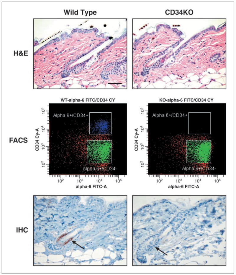
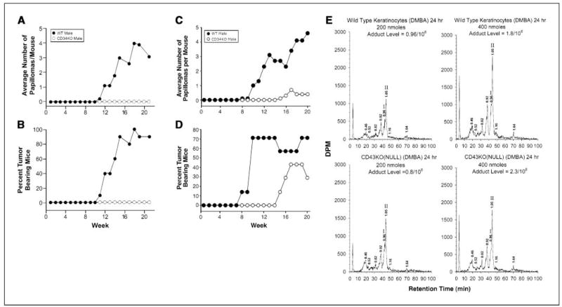
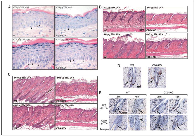
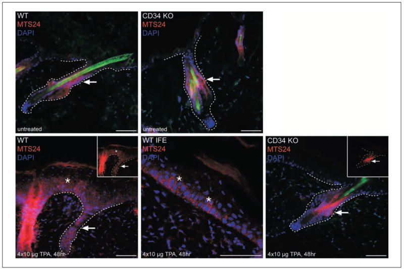
The cell surface marker CD34 marks mouse hair follicle bulge cells, which have attributes of stem cells, including quiescence and multipotency. Using a CD34 knockout (KO) mouse, we tested the hypothesis that CD34 may participate in tumor development in mice because hair follicle stem cells are thought to be a major target of carcinogens in the two-stage model of mouse skin carcinogenesis. Following initiation with 200 nmol 7,12-dimethylbenz(a)anthracene (DMBA), mice were promoted with 12-O-tetradecanoylphorbol-13-acetate (TPA) for 20 weeks. Under these conditions, CD34KO mice failed to develop papillomas. Increasing the initiating dose of DMBA to 400 nmol resulted in tumor development in the CD34KO mice, albeit with an increased latency and lower tumor yield compared with the wild-type (WT) strain. DNA adduct analysis of keratinocytes from DMBA-initiated CD34KO mice revealed that DMBA was metabolically activated into carcinogenic diol epoxides at both 200 and 400 nmol. Chronic exposure to TPA revealed that CD34KO skin developed and sustained epidermal hyperplasia. However, CD34KO hair follicles typically remained in telogen rather than transitioning into anagen growth, confirmed by retention of bromodeoxyuridine-labeled bulge stem cells within the hair follicle. Unique localization of the hair follicle progenitor cell marker MTS24 was found in interfollicular basal cells in TPA-treated WT mice, whereas staining remained restricted to the hair follicles of CD34KO mice, suggesting that progenitor cells migrate into epidermis differently between strains. These data show that CD34 is required for TPA-induced hair follicle stem cell activation and tumor formation in mice.


Figures

Figure 1
Figure 1
CD34KO mouse skin lacks CD34 expression and appears normal histologically. Top, skin from 7-wk-old WT and CD34KO mice was fixed in 10% formalin and stained with H & E; middle, single-cell suspensions of keratinocytes were stained with antibodies to α6 integrin and CD34 and analyzed by FACS; bottom, formalin-fixed sections were stained with a rat anti-mouse CD34 antibody. Bar, 50 μm.
Figure 2
Figure 2
Papilloma development in DMBA-initiated, TPA-promoted CD34KO mice. Seven-week-old CD34KO and WT mice were first initiated with subtumorigenic doses of DMBA and then promoted for 20 weeks with three applications per week of TPA in the standard two-stage mouse skin carcinogenesis regimen. Papillomas were counted weekly. A and B, mice were initiated with 200 nmol DMBA and then promoted thrice weekly with 4 μg TPA for 20 wks. A, average number of tumors per mouse. B, incidence of papillomas. C and D, CD34KO and WT mice were initiated with 400 nmol DMBA and then promoted thrice weekly with 5 μg TPA for 20 wks. C, average number of tumors per mouse. D, incidence of papillomas. E, mice were initiated with either 200 or 400 nmol DMBA, and keratinocytes were harvested 20 h later for DNA isolation and 32P-postlabeling identification of DMBADE DNA adducts. Top, DNA adducts for WT at 200 and 400 nmol; bottom, DNA adducts for CD34KO mice at 200 and 400 nmol. Syn-dAdo and anti-dAdo adducts are identified as I (retention time, 0.96 min) and II (retention time, 1.05 min), respectively.
Figure 3
Figure 3
Response of CD34KO epidermis and hair follicles to short-term TPA exposure. To test the ability of CD34KO skin to develop epidermal hyperplasia, 7-wk-old WT and CD34KO mice were subjected to short-term TPA exposure. Mice were dosed twice weekly for 2 wks with either 5 or 10 μg of TPA, and tissues were collected at 24 and 48 h after the last dose. In addition, beginning at postnatal day 3, CD34KO and WT pups were injected with BrdUrd twice daily for 3 d to generate LRCs. Tissues were fixed in formalin, sectioned, and stained with anti-BrdUrd to identify the quiescent LRCs both under steady-state conditions and after TPA treatment. A, H&E-stained, high-magnification photomicrographs of the interfollicular epidermis in WT (left) and CD34KO (right) to both doses of TPA, 48 h only. Bar, 20 μm. Arrowheads, basal cells. Compare the simple cuboidal cells of the CD34KO mouse with the irregular layers of larger elongated basal cells of WT mice. B and C, photomicrographs of H&E-stained skin taken 24 and 48 h after the last dose of either 4 × 5 μg TPA or 4 × 10 μg TPA. Bar, 100 μm. In WT mice, previously resting hair follicles enter the growing stage in response to TPA treatment. In contrast, hair follicles in CD34KO mice remain in telogen, the resting phase of the hair cycle. Left, WT images; right, CD34KO images. Compare the small inactive hair follicles and thin epidermis of CD34KO mice with the large active growing hair follicles extending deep into the thick dermis of WT mice. D, LRC localization in WT and CD34KO hair follicles after a 7-wk chase. Bar, 25 μm. E, LRC response after TPA treatment. Left, WT; right, CD34KO. Bar, 25 μm. Arrows, bulge. Note in (E) the highly cellular, active hair follicles in WT mice.
Figure 4
Figure 4
Localization of the hair follicle progenitor cell marker MTS24 in WT and CD34KO skin. To determine if lack of CD34 expression in hair follicles affected the localization of the hair follicle progenitor marker, untreated and TPA-treated CD34KO and WT skin was subjected to immunostaining with a mouse-specific MTS24 antibody. Top, localization to the midportion of telogen hair follicles (arrows) in untreated WT and CD34KO skin (7 wks of age); bottom, staining in anagen hair follicles (arrow) 48 h after the last of four applications of TPA (10 μg/dose). Note staining in the interfollicular epidermis extending into the suprabasal layers (*). Bottom middle, higher-magnification view of MTS24 labeling in basal cells of the interfollicular epidermis in WT skin after TPA (*); bottom right, MTS24 staining remains restricted to hair follicles in CD34KO (arrows) after TPA treatment. Insets, MTS24 staining (red) without 4′,6-diamidino-2-phenylindole (DAPI; blue). Bar, 50 μm.

References

    1. Cotsarelis G, Sun TT, Lavker RM. Label-retaining cells reside in the bulge area of pilosebaceous unit: implications for follicular stem cells, hair cycle, and skin carcinogenesis. Cell. 1990;61:1329–37. - PubMed
    1. Fuchs E, Tumbar T, Guasch G. Socializing with the neighbors: stem cells and their niche. Cell. 2004;116:769–78. - PubMed
    1. Lavker RM, Sun TT. Epidermal stem cells: properties, markers, and location. Proc Natl Acad Sci U S A. 2000;97:13473–5. - PMC - PubMed
    1. Morris RJ, Liu Y, Marles L, et al. Capturing and profiling adult hair follicle stem cells. Nat Biotechnol. 2004;22:411–7. - PubMed
    1. Claudinot S, Nicolas M, Oshima H, Rochat A, Barrandon Y. Long-term renewal of hair follicles from clonogenic multipotent stem cells. Proc Natl Acad Sci U S A. 2005;102:14677–82. - PMC - PubMed

Publication types

Substances