. 2007 May 1;67(9):4254-63.
doi: 10.1158/0008-5472.CAN-06-4100.

Angiopoietin-2 stimulates breast cancer metastasis through the alpha(5)beta(1) integrin-mediated pathway

Affiliations

Angiopoietin-2 stimulates breast cancer metastasis through the alpha(5)beta(1) integrin-mediated pathway

Yorihisha Imanishi et al. Cancer Res. .

Abstract

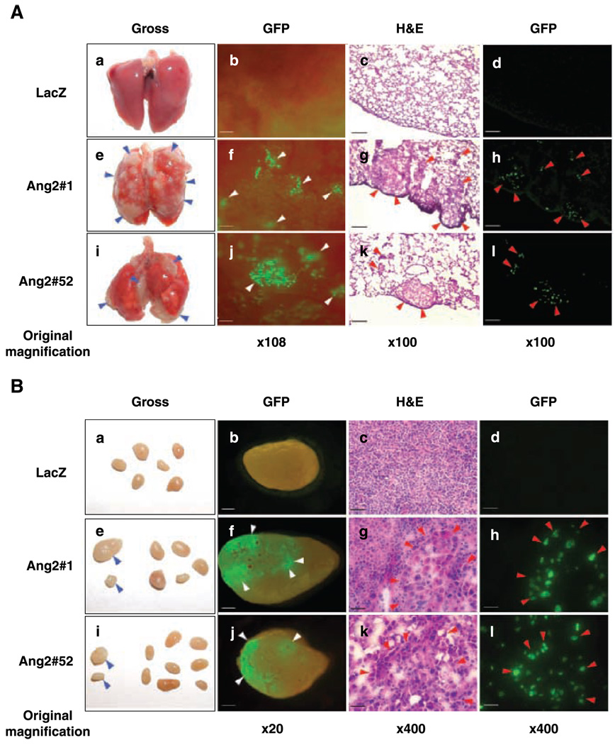
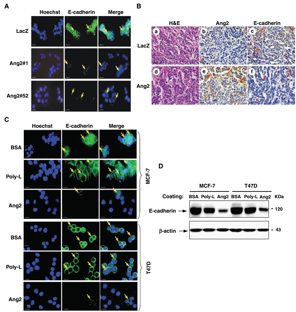
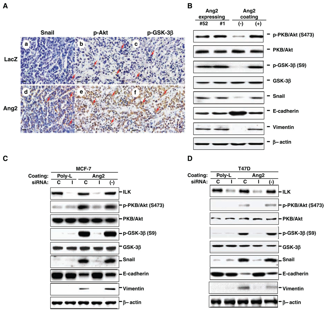
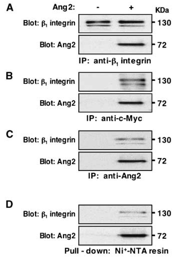
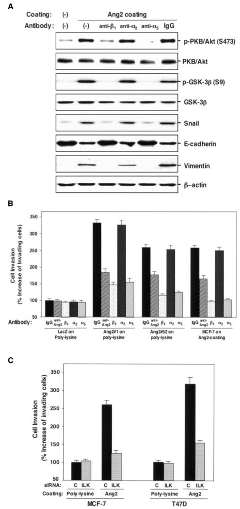
Acquisition of a metastatic phenotype by breast cancer cells includes alternations of multigenic programs that permit tumor cells to metastasize to distant organs. Here, we report that angiopoietin-2 (Ang2), a known growth factor, is capable of promoting breast cancer cell invasion leading to metastasis. Analysis of 185 primary human breast cancer specimens that include 97 tumors showing lymph node and/or distant metastasis reveals a significant correlation between the expression of Ang2 and E-cadherin, Snail, metastatic potential, tumor grade, and lymph-vascular invasion during breast cancer progression. Using a xenograft model, we show that overexpression of Ang2 in poorly metastatic MCF-7 breast cancer cells suppresses expression of E-cadherin and induces Snail expression and phosphorylation of Akt and glycogen synthase kinase-3beta (GSK-3beta) promoting metastasis to the lymph nodes and lung. In cell culture, Ang2 promotes cell migration and invasion in Tie2-deficient breast cancer cells through the alpha(5)beta(1) integrin/integrin-linked kinase (ILK)/Akt, GSK-3beta/Snail/E-cadherin signaling pathway. Inhibition of ILK and the alpha(5)beta(1) integrin abrogates Ang2 modulation of Akt, GSK-3beta, Snail, and E-cadherin and Ang2-stimulated breast cancer cell migration and invasion. Together, these results underscore the significant contribution of Ang2 in cancer progression, not only by stimulating angiogenesis but also by promoting metastasis, and provide a mechanism by which breast cancer cells acquire an enhanced invasive phenotype contributing to metastasis.


Figures

Figure 1
Figure 1
Expression of Ang2 correlates with Snail and E-cadherin expression and metastatic potential in primary human breast cancer specimens. A, human breast cancer specimens were stained for Ang2 (a and b), Snail (b and e), and E-cadherin (c and f). Arrows, positive staining of Ang2 (a and d), Snail (b and e, especially in the nuclei), or E-cadherin (c, dominantly on the cell membrane; f , barely positive in the cytoplasm) in the tumor cells. Ang2 proteins were detected mostly in tumor cell cytoplasm. Representative stains from a total of 185 tumor samples. Each specimen was independently analyzed at least twice with similar results. Bar, 20 µm. B, expression of Ang2 mRNA correlates with the metastatic potential of primary human breast cancer specimens. Quantitative real-time PCR was done on cDNA derived from total RNA isolated from nonmetastatic and metastatic snap-frozen human breast cancer specimens. The relative expression of Ang2 in the metastatic versus the nonmetastatic samples was calculated using a difference in threshold cycle (ΔCt) method with β-glucuronidase as the normalizing control gene. PCRs were done in triplicate and each specimen was independently analyzed twice with similar results.
Figure 2
Figure 2
Overexpression of Ang2 by MCF-7 breast cancer cells promotes tumor metastasis in mice. MCF-7 parental cells were obtained from ATCC. Various MCF-7 cell lines were generated to stably express Ang2 or LacZ and GFP with no observable alterations in cell properties (see Materials and Methods in the Supplementary Data). A and B, tumor metastasis was examined by gross observation (a, e, and i) and epifluorescent observation of GFP (b, f , and j). Cryosections were further examined by epifluorescence (d, h, and l) followed by H&E staining (c, g, and k). Mice that received MCF-7/Ang2/GFP cells (clone 1 or 52) showed metastasis in the lung and lymph nodes (e–l in A and B) with high incidence. Grossly visible metastases (blue arrowheads) in the lung (e and i in A) and lymph nodes (e and i in B) were identified as green foci (f and j, white arrowheads) and micrometastatic cells expressing GFP (h and l, red arrowheads) and by H&E staining (g and k , red arrowheads). Bars, 120 µm (b, c, d, f, g, h, j, k, and l in A), 600 µm (b, f , and j in B), and 30 µm (c, d, g, h, k, and l in B). The experiments in (A) and (B) were done three independent times and similar results were obtained. Additionally, these experiments were also done twice using Ang2#4 (low expression of Ang2) and Ang2#5 (high expression of Ang2; Supplementary Fig. S1A) in mice. High incidence of lymph node and lung metastasis was found in mice that received Ang2#5 cells, whereas no tumor metastasis was found in mice that received Ang2#4 cells.
Figure 3
Figure 3
Ang2 stimulation induces loss of E-cadherin expression in breast cancer cells. MCF-7 and T47D cells used in Fig. 3 to Fig 6 were obtained from ATCC. A, immunofluorescent staining of MCF-7/LacZ control and MCF-7/Ang2 cells (clones 1 and 52) using an anti-E-cadherin antibody (green) and Hoechst (blue for nuclear staining). MCF-7/Ang2 cells showed decreased expression of E-cadherin in the cell membrane leading to loss of cell-cell contact (arrows in MCF-7/Ang2 cells). Bar, 15 µm. B, immunohistochemical analyses of orthotopic tumors established by LacZ/GFP (a–c) or Ang2#1 cells (d and f) using H&E (a and c), anti-Ang2 (b and e), or anti-E-cadherin antibodies (c and f). Arrows, Ang2 (b and e) or E-cadherin (c and f) staining. Three to five serial-cut paraffin sections from five or more individual tumor samples of each group were independently analyzed. Bars, 20 µm. C, immunofluorescent staining of MCF-7 and T47D cells adhered to exogenous Ang2, heat-inactivated BSA, or poly-lysine (Poly-L). Down-regulation of E-cadherin in the cell membrane and loss of cell-cell contact (arrows) were observed in Ang2-treated cells. Bar, 15 µm. D, Western blot analyses of E-cadherin expression in exogenous Ang2-treated breast cancer cells. MCF-7 and T47D cells were treated with heat-inactivated BSA, Ang2, or poly-lysine for 48 h. β-Actin was used as a loading control. The experiments in (A) to (D) were done two independent times with similar results.
Figure 4
Figure 4
Ang2 stimulates the ILK/Akt, GSK-3β/Snail/E-cadherin pathway. A, immunohistochemical analyses of identical orthotopic tumors established by LacZ/GFP (a–c) or Ang2#1 (d–f) using anti-Snail antibody (a and d, paraffin-embedded tissue sections), anti-p-Akt (Ser473) antibody (b and e, frozen tissue sections), and anti-p-GSK-3β (Ser9) antibody (c and f, frozen tissue sections). Arrows, positive staining of Snail especially in nuclei of tumor cells (a and d), p-Akt in cytoplasm (b and e), or p-GSK-3β in cytoplasm (c and f). Three to five serial-cut sections from five or more individual tumor samples of each group were independently analyzed. Bar, 20 µm. B, Western blot analyses of Ang2-expressing cells or LacZ control cells treated with Ang2 using anti-p-Akt (S473), anti-p-GSK-3β (S9), anti-Snail, anti-E-cadherin, and anti-vimentin antibodies. The membranes were reprobed with anti-Akt, anti-GSK-3β, or anti-β-actin antibodies as loading controls. C and D, Western blot analyses of parental MCF-7 (C) and T47D (D) cells transfected with siRNA for ILK (I), control siRNA (C), or no transfection (−) followed by stimulation with Ang2 or poly-lysine. The cell lysates were then analyzed as described in (B) except an anti-ILK antibody was used. The results from (A) to (D) are representative of three independently repeated experiments.
Figure 5
Figure 5
Ang2 associates with β1 integrin in Tie2-deficient MCF-7 cells. Coimmunoprecipitation (IP) and pull-down assays. Exogenous Ang2 was encoded by a c-Myc and His-tagged pSecTagB expression vector that is described in the Supplementary Data and expressed in MCF-7 Ang2#1 cells. The associated Ang2 with β1 integrin on various cells was assessed by immunoprecipitation separately with anti-β1 (A), anti-c-Myc (B), or anti-Ang2 (C) antibodies followed by Western blot using anti-β1 (A–C, top) or anti-Ang2 (A–C, bottom) antibodies. D, a pull-down assay. The Ang2-associated β1 integrin was pulled down by its His tag of Ang2 using Ni+-NTA beads followed by Western blot using anti-β1 or anti-Ang2 antibodies. Similar results were also found when the identical experiments were done using T47D cells (data not shown). Results are representative of three independently repeated experiments.
Figure 6
Figure 6
Ang2-stimulated breast cancer cell invasion is mediated through the α5β1 integrin-mediated pathway. A, Western blot analyses. Inhibition of β1 and α5, but not α2, integrins attenuates Ang2 modulation of Akt, GSK-3β, Snail, E-cadherin, and vimentin. The membranes were reprobed with anti-Akt, anti-GSK-3β, or anti-β-actin antibodies as loading controls. B and C, in vitro cell invasion assays. B, inhibition of β1 and α5, but not α2, integrins suppresses Ang2 stimulation of breast cancer cell invasion. Mouse IgG and an anti-Ang2 (a nonneutralizing) antibody are included as controls. C, inhibition of ILK using siRNA specific for ILK suppresses Ang2 stimulation of breast cancer cell invasion. Results in (A) to (C) are representative of three independent times. Columns, mean percentage increase in the number of invading cells compared with control; bars, SD.

References

    1. Chambers AF, Groom AC, MacDonald IC. Dissemination and growth of cancer cells in metastatic sites. Nat Rev Cancer. 2002;2:563–572. - PubMed
    1. Thiery JP. Epithelial-mesenchymal transitions in tumour progression. Nat Rev Cancer. 2002;2:442–454. - PubMed
    1. Nieto MA. The snail superfamily of zinc-finger transcription factors. Nat Rev Mol Cell Biol. 2002;3:155–166. - PubMed
    1. Zhou BP, Deng J, Xia W, et al. Dual regulation of Snail b y GSK-3β-mediated phosphorylation in control of epithelial-mesenchymal transition. Nat Cell Biol. 2004;6:931–940. - PubMed
    1. Barbera MJ, Puig I, Dominguez D, et al. Regulation of Snail transcription during epithelial to mesenchymal transition of tumor cells. Oncogene. 2004;23:7345–7354. - PubMed

Publication types

MeSH terms