. 2007 Jul;176(3):1541-55.
doi: 10.1534/genetics.107.073494. Epub 2007 May 4.

Phosphorylation of the Sic1 inhibitor of B-type cyclins in Saccharomyces cerevisiae is not essential but contributes to cell cycle robustness

Affiliations

Phosphorylation of the Sic1 inhibitor of B-type cyclins in Saccharomyces cerevisiae is not essential but contributes to cell cycle robustness

Frederick R Cross et al. Genetics. 2007 Jul.

Abstract

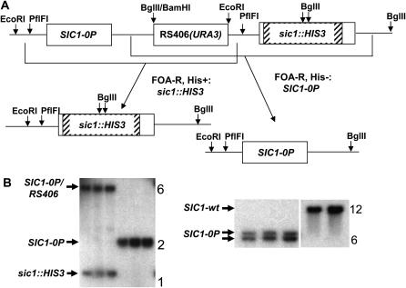
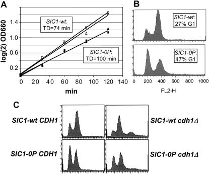
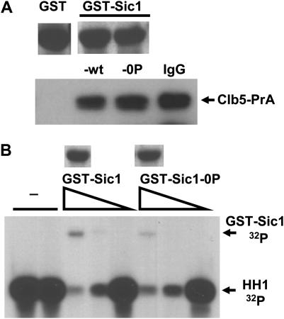
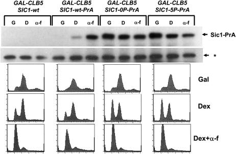
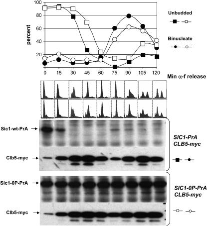
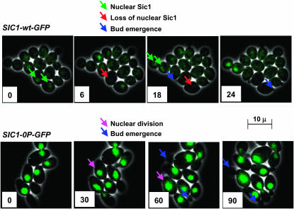
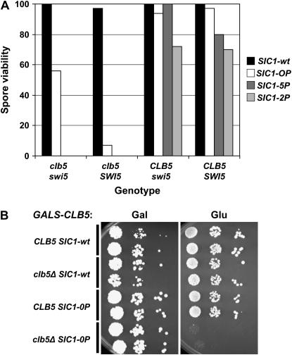
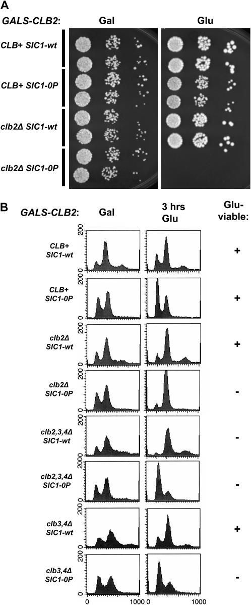
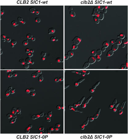
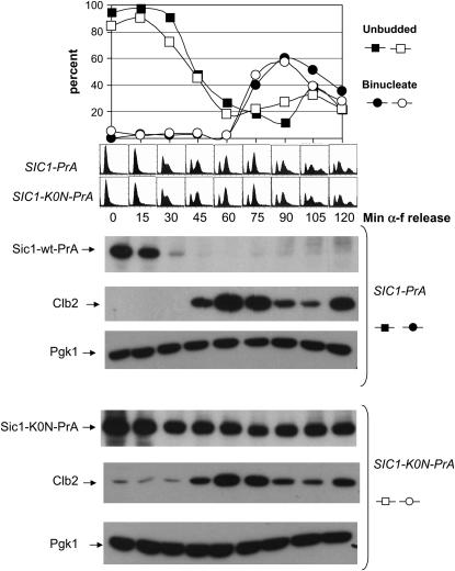
In budding yeast, B-type cyclin (Clb)-dependent kinase activity is essential for S phase and mitosis. In newborn G(1) cells, Clb kinase accumulation is blocked, in part because of the Sic1 stoichiometric inhibitor. Previous results strongly suggested that G(1) cyclin-dependent Sic1 phosphorylation, and its consequent degradation, is essential for S phase. However, cells containing a precise endogenous gene replacement of SIC1 with SIC1-0P (all nine phosphorylation sites mutated) were fully viable. Unphosphorylatable Sic1 was abundant and nuclear throughout the cell cycle and effectively inhibited Clb kinase in vitro. SIC1-0P cells had a lengthened G(1) and increased G(1) cyclin transcriptional activation and variable delays in the budded part of the cell cycle. SIC1-0P was lethal when combined with deletion of CLB2, CLB3, or CLB5, the major B-type cyclins. Sic1 phosphorylation provides a sharp link between G(1) cyclin activation and Clb kinase activation, but failure of Sic1 phosphorylation and proteolysis imposes a variable cell cycle delay and extreme sensitivity to B-type cyclin dosage, rather than a lethal cell cycle block.


Figures

F<sc>igure</sc> 1.—
Figure 1.—
Scheme for replacement of SIC1 with SIC1-0P. (A) SIC1-0P, including a 5′ promoter sequence, in the cloning vector RS406 (URA3) (plasmid FC663) was digested with PflFI, cutting in the SIC1 promoter, and used to transform a sic1∷HIS3 strain. The expected resulting structure is shown on top (not to scale). Homologous recombination events resulting in Ura3− popout derivatives were selected on the basis of 5-FOA resistance and were found to be a mix of His+ and His−. These were interpreted as being due to recombination in the regions indicated by brackets. (B) Selected Southern blot analysis: (Left) EcoRI+BglII digestion of DNA from three integrants of RS406-SIC1-0P at the sic1∷HIS3 locus (first three lanes) and FOA-resistant His− popouts derived from these integrants (second three lanes), probed with SIC1 DNA from EcoRI to KpnI (at border of HIS3 insertion in sic1∷HIS3), confirming replacement of sic1∷HIS3 with SIC1-0P in the His− popouts. (Right) DNA from the same three FOA-resistant His− popouts and two SIC1-wt controls digested with ApaI and probed with a SIC1 coding sequence probe, confirming an ApaI site introduced at the S80A phosphorylation-site mutation (near the center of an ∼12-kb genomic ApaI fragment containing SIC1). Deduced identities of the bands are indicated; band sizes are approximately as expected from known genomic sequence.
F<sc>igure</sc> 2.—
Figure 2.—
SIC1-0P cells are viable with an elongated G1. (A) SIC1-0P SWI5 and SIC1-wt SWI5 strains were constructed and grown in YEPD at 30°. At intervals, the OD660 was determined to measure the doubling times, indicated in the insets. (B) In the middle of the growth curve shown in A, samples were taken for flow-cytometry analysis. An approximate quantitation of the proportion of 1C DNA content (G1) cells was made (insets). (C) A SIC1-0P strain was crossed to a cdh1∷LEU2 sic1∷HIS3 pURA3-SIC1 strain, and tetrads were dissected. Flow-cytometry data from representative segregants (all lacking the pURA3-SIC1 plasmid) are presented.
F<sc>igure</sc> 3.—
Figure 3.—
Sic1-0P binds and inhibits Clb5-Cdc28. (A) GST, GST-Sic1, and GST-Sic1-0P were purified from E. coli on glutathione–sepharose beads. VAY79 (cdc20 GAL-CDC20 CLB5-PrA) was blocked in glucose medium to deplete Cdc20 and allow accumulation of high levels of Clb5-PrA. Extracts made from this culture were incubated for 1 hr on ice with glutathione–sepharose beads containing equivalent amounts of GST, GST-Sic1, or GST-Sic1-0P (parallel amido black stains for GST fusions at top of figure) or with IgG–sepharose beads to directly purify Clb5 via the PrA tag, and beads were washed by repeated centrifugation. Western blot analysis of bead-bound Clb5-PrA is shown. (B) Clb5-PrA was purified from cell extracts as in A using IgG–sepharose. Aliquots (15 μl) of Clb5-PrA–IgG–agarose bead suspension in kinase buffer were incubated for 15 min on ice with no addition, with 1 μl of glutathione elution buffer, or with glutathione elution buffer containing 10-fold serial dilutions of soluble GST-Sic1 fusions (an estimated addition of ∼600, 60, and 6 ng of GST-Sic1 or GST-Sic1-0P; parallel amido black stains of GST fusions at top). Then 5 μm ATP, 50 μCi 32P-labeled ATP, and 2 μg histone H1 were added (final 21 μl). Reactions were incubated for 10 min at 30°, stopped with SDS sample buffer, and separated by gel electrophoresis before exposure to film to assess histone H1 kinase activity. Phosphorylation of GST-Sic1, and less efficient phosphorylation of GST-Sic1-0P, were also detected, as indicated.
F<sc>igure</sc> 4.—
Figure 4.—
Sic1-0P and Sic1-5P fail to exhibit regulated changes in abundance displayed by wild-type Sic1. Strains carrying SIC1-wt, SIC1-0P, or SIC1-5P, all fused to a C-terminal protein A tag and expressed from the endogenous locus and also carrying a GALS-CLB5 cassette, were grown in galactose medium to overexpress Clb5 (G), shifted to glucose medium for 2.5 hr to shut off GALS-CLB5 (D), or to glucose medium plus α-factor (α-f) for 2.5 hr to arrest cells in G1. (Top) Western blots to detect Sic1-PrA or a control cross-reacting band (*) as loading control. Flow-cytometry profiles for the samples are presented below.
F<sc>igure</sc> 5.—
Figure 5.—
Sic1-0P is stable throughout the cell cycle. SIC1-wt-PrA or SIC1-0P-PrA GALS-CLB5 myc-CLB5 strains pregrown in galactose medium were switched to glucose medium plus α-factor (α-f) to arrest cells in G1, as in Figure 4. After a 2.5 hr arrest, cells were centrifuged, washed twice to remove α-factor, and reinoculated into fresh glucose medium. Time points were taken every 15 min. (Bottom) Western blot analysis of Sic1-PrA and myc-Clb5. The proportions of unbudded and binucleate cells were determined microscopically (top), and the approximate period of DNA replication (between 15 and 45 min after release) was determined by FACS (middle).
F<sc>igure</sc> 6.—
Figure 6.—
Nuclear Sic1-wt-GFP is detected only during M/G1; Sic1-0P-GFP is nuclear throughout the cell cycle. Cells containing SIC1-wt-GFP or SIC1-0P-GFP, expressed from the endogenous locus were analyzed by time-lapse fluorescence microscopy, with illumination every 3 min. Selected frames from movies are shown to illustrate Sic1-wt-GFP appearing as a nuclear signal in large-budded cells (top, green arrows) and then disappearing rapidly from the mother-cell nucleus and more slowly from the daughter-cell nucleus (top, red arrows). Sic1-wt-GFP disappearance occurs shortly before bud emergence (top, blue arrows). Sic1-0P-GFP was nuclear throughout the cell cycle, including before and after bud emergence (bottom, blue arrows) and before and after nuclear division (pink arrows). The disappearance of Sic1-0P-GFP from nuclei was never observed. In both series, for clarity, only some events are labeled with arrows.
F<sc>igure</sc> 7.—
Figure 7.—
Analysis of the cell cycle in SIC1-0P cells by quantitative time-lapse fluorescence microscopy using a CLN2pr-GFP marker. Time-lapse fluorescence microscopy on SIC1-wt or SIC1-0P cells also containing CLN2pr-GFP (unstable GFP under control of the CLN2 promoter) (Mateus and Avery 2000; Bean et al. 2006) was carried out as described (Bean et al. 2006) with 3-min resolution. (Top) Composite phase/fluorescence images from representative movies are presented [images taken every 15 min, starting 60 min (SIC1-wt) or 101 min (SIC1-0P) after plating]. The images were collected in parallel and processed identically. Background fluorescence corresponding to autofluorescence in unlabeled cells was subtracted. Graphs (middle) present data extracted automatically from all data collected (annotated files for microcolonies developed over 9 hr of exponential growth from seven mutant and six wild-type founder cells). The traces (left) represent the smoothed and background-subtracted CLN2pr-GFP intensity profiles for all cases where the peak finder found two successive peaks on a common scale for wild type and SIC1-0P. The histogram (bottom) indicates the distribution of times between successive mother-cell buddings, measuring the mother-cell cycle time.
F<sc>igure</sc> 8.—
Figure 8.—
Phosphorylation-site mutations in SIC1 result in lethality in the absence of CLB5. (A) Tetrad analysis was performed on diploids of genotypes swi5∷kanMX/+ clb5∷URA3/+, sic1∷HIS3/SIC1-wt, sic1∷HIS3/SIC1-0P, sic1∷HIS3/SIC1-5P, or sic1∷HIS3/SIC1-2P: SIC1-5P, T2A, T5A, T33A, S76A; SIC1-2P, T33A, S69A, S76A, T80A, S174A, NS T192A. Spore viability for various genotypes was assessed assuming 2:2 segregation for all markers. Rare viable segregants of the genotype clb5 SWI5 SIC1-0P were small, slow-growing colonies. (B) GALS-CLB5 SIC1-0P clb5∷URA3 strains and controls were constructed by tetrad analysis on galactose medium. Serial dilutions on galactose medium (Gal; GALS-CLB5 on) and glucose medium (Glu; GALS-CLB5 off) were incubated for 3 days at 30°.
F<sc>igure</sc> 9.—
Figure 9.—
Requirements for mitotic CLB2–4 in a SIC1-0P background. (A) GALS-CLB2 SIC1-0P clb2∷LEU2 strains and controls were constructed by tetrad analysis on galactose medium. Serial dilutions on galactose medium (Gal; GALS-CLB on) and glucose medium (Glu; GALS-CLB off) were incubated for 3 days at 30°. (B) Flow-cytometry analysis of GALS-CLB2 strains, either SIC1-wt or SIC1-0P, containing the indicated additional CLB gene deletions. Strains were grown in galactose medium (Gal) or transferred to glucose medium (Glu) for 3 hr. (Right) Viability of the indicated genotype on glucose medium (GALS-CLB2 off) as determined by a replica-plating patch assay.
F<sc>igure</sc> 10.—
Figure 10.—
Lethal phenotype of clb2 SIC1-0P cells. Cells of the indicated genotype, also containing a GALS-CLB2 cassette, were grown in galactose medium to log phase and transferred to glucose medium for 3 hr. Cells were fixed in ethanol and digested with RNase and protease and nuclear DNA was stained with propidium iodide; samples were examined by DIC and fluorescence microscopy, and composite images were generated. All exposure settings were identical for these images.
F<sc>igure</sc> 11.—
Figure 11.—
Sic1-K0N is relatively stable throughout the cell cycle. SIC1-wt-PrA or SIC1-K0N-PrA strains were grown to log phase in glucose medium and then α-factor (α-f) was added to arrest cells in G1, as in Figure 4. After a 2.5 hr arrest, cells were centrifuged and washed twice to remove α-factor and reinoculated into fresh glucose medium. Time points were taken every 15 min. (Bottom) Western blot analysis of Sic1-PrA, Clb2, and Pgk1 as a loading control. The proportions of unbudded and binucleate cells were determined microscopically, and the approximate period of DNA replication (between 15 and 45 min after release) by flow cytometry (middle panels).

Similar articles

Cited by

References

    1. Amon, A., M. Tyers, B. Futcher and K. Nasmyth, 1993. Mechanisms that help the yeast cell cycle clock tick: G2 cyclins transcriptionally activate G2 cyclins and repress G1 cyclins. Cell 74: 993–1007. - PubMed
    1. Bar-Even, A., J. Paulsson, N. Maheshri, M. Carmi, E. O'Shea et al., 2006. Noise in protein expression scales with natural protein abundance. Nat. Genet. 38: 636–643. - PubMed
    1. Bean, J. M., E. D. Siggia and F. R. Cross, 2006. Coherence and timing of cell cycle Start examined at single-cell resolution. Mol. Cell 21: 3–14. - PubMed
    1. Bi, E., P. Maddox, D. J. Lew, E. D. Salmon, J. N. McMillan et al., 1998. Involvement of an actomyosin contractile ring in Saccharomyces cerevisiae cytokinesis. J. Cell Biol. 142: 1301–1312. - PMC - PubMed
    1. Chen, K. C., L. Calzone, A. Csikasz-Nagy, F. R. Cross, B. Novak et al., 2004. Integrative analysis of cell cycle control in budding yeast. Mol. Biol. Cell 15: 3841–3862. - PMC - PubMed

Publication types

MeSH terms