. 2007 Jul;176(3):1417-29.
doi: 10.1534/genetics.107.072876. Epub 2007 May 4.

Genetic dissection of parallel sister-chromatid cohesion pathways

Affiliations

Genetic dissection of parallel sister-chromatid cohesion pathways

Hong Xu et al. Genetics. 2007 Jul.

Abstract

Sister-chromatid cohesion, the process of pairing replicated chromosomes during mitosis and meiosis, is mediated through the essential cohesin complex and a number of nonessential cohesion genes, but the specific roles of these nonessential genes in sister-chromatid cohesion remain to be clarified. We analyzed sister-chromatid cohesion in double mutants of mrc1Delta, tof1Delta, and csm3Delta and identified additive cohesion defects that indicated the existence of at least two pathways that contribute to sister-chromatid cohesion. To understand the relationship of other nonessential cohesion genes with respect to these two pathways, pairwise combinations of deletion and temperature-sensitive alleles were tested for cohesion defects. These data defined two cohesion pathways, one containing CSM3, TOF1, CTF4, and CHL1, and the second containing MRC1, CTF18, CTF8, and DCC1. Furthermore, we found that the nonessential genes are not important for the maintenance of cohesion at G(2)/M. Thus, our data suggest that nonessential cohesion genes make critical redundant contributions to the establishment of sister-chromatid cohesion and define two cohesion pathways, thereby establishing a framework for understanding the role of nonessential genes in sister-chromatid cohesion.


Figures
F<sc>igure</sc> 1.—
Figure 1.—
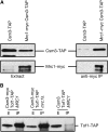
Mrc1 physically interacts with Csm3. (A) Extracts from yeast strains expressing the indicated epitope-tagged proteins were immunoprecipitated with anti-myc antibodies followed by protein G agarose. Ten percent of the input extract and 50% of the immunoprecipitate were fractionated by SDS–PAGE. Immunoblots were probed with peroxidase–antiperoxidase to detect Csm3-TAP and with α-myc to detect Mrc1-myc. (B) Extracts from MRC1 or mrc1Δ yeast strains expressing Csm3-myc and Tof1-TAP were immunoprecipitated with anti-myc antibodies followed by protein G agarose. Ten percent of the input extract and 50% of the immunoprecipitate were fractionated by SDS–PAGE. Immunoblots were probed with peroxidase–antiperoxidase to detect Tof1-TAP.
F<sc>igure</sc> 2.—
Figure 2.—
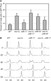
mrc1Δ tof1Δ and mrc1Δ csm3Δ exhibit additive cohesion defects relative to single mutants. (A) Micrographs of mrc1Δ tof1Δ in which one sister-chromatid locus was visualized with TetR-GFP. (Top) DIC image. (Bottom) Fluorescence image. (B) Analysis of cohesion in wild-type and mutant strains at 30° in nocodazole-arrested cells. The relevant genotypes are indicated, and the number of GFP signals in each cell was scored for at least two independent strains in independent experiments. At least 200 cells were scored for each strain. (C) The percentage of cells with Pds1 signal was determined for all cells, and for cells with two GFP foci. Pds1-18myc protein was detected by indirect immunofluorescence. (D–F) Analysis of cohesion in wild-type (D), mrc1Δtof1Δ (E), and mrc1Δcsm3Δ (F) strains without nocodazole arrest. Cells were blocked in G1 phase by incubating with α-factor for 3 hr before synchronous release into the cell cycle. Samples were taken every 20 min and analyzed for the presence of paired or separated GFP foci and for the presence of Pds1.
F<sc>igure</sc> 3.—
Figure 3.—
mrc1Δ tof1Δ does not exhibit additive replication or checkpoint defects relative to the single mutants. (A) Flow cytometric analysis of DNA contents during synchronous progression through S phase. Log-phase cultures were synchronized in G1 with α-factor and then released into YPD medium at 37°. Samples were collected at the indicated times and DNA contents were analyzed by flow cytometry. The relevant genotypes are indicated, as are the positions of cells with 1C and 2C DNA contents. (B) Rad53 activation. Log-phase cultures of the indicated strains were treated with 0.035% MMS for 1 hr (+) or were mock treated (−). Samples were analyzed on immunoblots. The positions of Rad53 and the activated, phosphorylated form of Rad53 (Rad53-P) are indicated.
F<sc>igure</sc> 4.—
Figure 4.—
csm3-9 ctf18Δ accumulates large-budded cells at the restrictive temperature. (A) DIC images of wild type (left) and csm3-9 ctf18Δ (right) after growth at the restrictive temperature (37°) for 4 hr. (B) Nuclear morphology of wild-type, csm3-9, ctf18Δ, and csm3-9 ctf18Δ strains was scored by staining with DAPI after 4 hr at the restrictive temperature (37°). Nuclear morphology was classified into five categories as indicated. (C) Flow cytometric histograms displaying DNA contents for the indicated strains during asynchronous growth at the permissive temperature. (D) Quantification of nuclear morphology for wild type, csm3-9 ctf18Δ, csm3-9 ctf18Δ mad2Δ, and csm3-9 ctf18Δ rad9Δ following growth at the restrictive temperature for 4 hr.
F<sc>igure</sc> 5.—
Figure 5.—
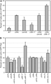
(A) csm3-9 exhibits additive cohesion defects when combined with mutants in ctf8, ctf18, or dcc1. Sister-chromatid cohesion assays were performed at both the permissive (26°) and the restrictive temperature (37°) in nocodazole-arrested cells. The relevant genotypes are indicated, and the number of GFP signals in each cell was scored for at least two independent strains in independent experiments. At least 200 cells were scored for each strain. (B) ctf8-17 has an additive cohesion defect with csm3Δ and tof1Δ. Cohesion assays were performed at the restrictive temperature (37°) following arrest in nocodazole. The number of GFP signals in each cell was scored for at least two independent strains in independent experiments and at least 200 cells were scored for each strain.
F<sc>igure</sc> 6.—
Figure 6.—
CTF8 is in the same cohesion pathway as MRC1. (A) Cohesion assays were performed at the restrictive temperature (37°) following treatment with nocodazole. The relevant genotypes are indicated, and the number of GFP signals in each cell was scored for at least two independent strains in independent experiments. At least 200 cells were scored for each strain. (B) S-phase progression was analyzed by flow cytometry. Log-phase cultures were synchronized in G1 with α-factor at 26° and then released into YPD medium at 37°. Samples were collected at the indicated times and DNA contents were analyzed by flow cytometry. The relevant genotypes are indicated, as are the positions of cells with 1C and 2C DNA contents.
F<sc>igure</sc> 7.—
Figure 7.—
Ctf4 and Chl1 are additional members of the Csm3/Tof1 cohesion pathway. (A) Ctf4 is in the same cohesion pathway as Csm3/Tof1. Cohesion assays were performed on the indicated strains, and the number of GFP signals in each cell was scored for at least two independent strains in independent experiments. At least 200 cells were scored for each strain. (B) Chl1 is in the same cohesion pathway as Csm3/Tof1, parallel with the Ctf8 pathway. Cohesion assays were performed as in A.
F<sc>igure</sc> 8.—
Figure 8.—
The nonessential cohesion genes are not required for maintenance of cohesion. The indicated strains, with Cdc20 expressed exclusively under the control of the GAL1-10 promoter, were grown in the presence of galactose (Cdc20 on) at 26° to mid-logarithmic phase and then transferred to glucose medium (Cdc20 off) for 3.5 hr to block cells in M phase. To test if each gene's function is required for cohesion maintenance, the temperature was shifted to 37° for 1.5 hr after blocking in M phase (solid bars). As controls, cell cultures were kept at 26° (open bars) following the M-phase block or were incubated at 37° (shaded bars) during the 3.5-hr M-phase block in glucose and after the M-phase block. The number of GFP signals in each cell was scored in at least two independent experiments. At least 200 cells were scored for each experiment.
F<sc>igure</sc> 9.—
Figure 9.—
Cohesion analysis correlates with genetic interactions between two cohesion pathways. Results of cohesion analysis are represented by straight blue lines. Solid blue lines indicate additive cohesion defects in double mutants; dashed blue lines indicate an absence of an additive cohesion defect in double mutants. Genetic interactions are represented by curved red lines. Solid red lines indicate synthetic-lethal interactions; dashed red lines indicate synthetic-sick interactions. Where there is no red line between two genes, a genetic interaction was not evident. The genetic interactions for each subunit of the Ctf18 complex, and for Tof1 and Csm3, are the same; therefore only one line is used to link each complex with other genes. Genetic interaction data are from Tong et al. (2004).

Similar articles

Cited by

References

    1. Alcasabas, A. A., A. J. Osborn, J. Bachant, F. Hu, P. J. Werler et al., 2001. Mrc1 transduces signals of DNA replication stress to activate Rad53. Nat. Cell Biol. 3: 958–965. - PubMed
    1. Baetz, K. K., N. J. Krogan, A. Emili, J. Greenblatt and P. Hieter, 2004. The ctf13–30/CTF13 genomic haploinsufficiency modifier screen identifies the yeast chromatin remodeling complex RSC, which is required for the establishment of sister chromatid cohesion. Mol. Cell. Biol. 24(3): 1232–1244. - PMC - PubMed
    1. Bailis, J. M., P. Bernard, R. Antonelli, R. C. Allshire and S. L. Forsburg, 2003. Hsk1-Dfp1 is required for heterochromatin-mediated cohesion at centromeres. Nat. Cell Biol. 5: 1111–1116. - PubMed
    1. Barbour, L., and W. Xiao, 2003. Regulation of alternative replication bypass pathways at stalled replication forks and its effects on genome stability: a yeast model. Mutat. Res. 532: 137–155. - PubMed
    1. Bellaoui, M., M. Chang, J. Ou, H. Xu, C. Boone et al., 2003. Elg1 forms an alternative RFC complex important for DNA replication and genome integrity. EMBO J. 22: 4304–4313. - PMC - PubMed

Publication types

MeSH terms

Substances