. 2007 Jul;176(3):1567-77.
doi: 10.1534/genetics.107.072587. Epub 2007 May 4.

Oxidative stress enzymes are required for DAF-16-mediated immunity due to generation of reactive oxygen species by Caenorhabditis elegans

Affiliations

Oxidative stress enzymes are required for DAF-16-mediated immunity due to generation of reactive oxygen species by Caenorhabditis elegans

Violeta Chávez et al. Genetics. 2007 Jul.

Abstract

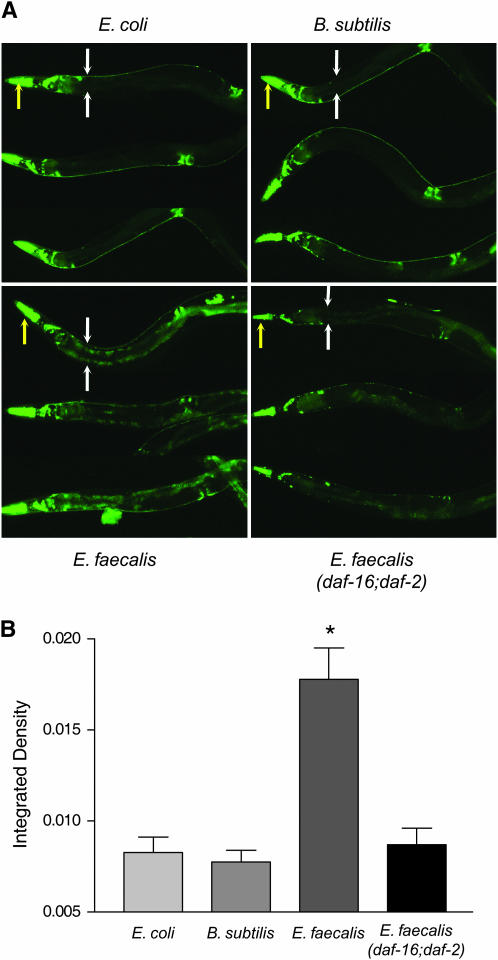
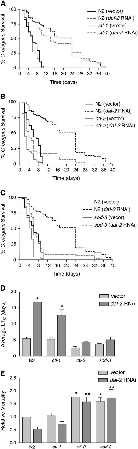
Caenorhabditis elegans has recently been developed as a model for microbial pathogenesis, yet little is known about its immunological defenses. Previous work implicated insulin signaling in mediating pathogen resistance in a manner dependent on the transcriptional regulator DAF-16, but the mechanism has not been elucidated. We present evidence that C. elegans, like mammalian phagocytes, produces reactive oxygen species (ROS) in response to pathogens. Signs of oxidative stress occur in the intestine - the site of the host-pathogen interface - suggesting that ROS release is localized to this tissue. Evidence includes the accumulation of lipofuscin, a pigment resulting from oxidative damage, at this site. In addition, SOD-3, a superoxide dismutase regulated by DAF-16, is induced in intestinal tissue after exposure to pathogenic bacteria. Moreover, we show that the oxidative stress response genes sod-3 and ctl-2 are required for DAF-16-mediated resistance to Enterococcus faecalis using a C. elegans killing assay. We propose a model whereby C. elegans responds to pathogens by producing ROS in the intestine while simultaneously inducing a DAF-16-dependent oxidative stress response to protect adjacent tissues. Because insulin-signaling mutants overproduce oxidative stress response enzymes, the model provides an explanation for their increased resistance to pathogens.


Figures

F<sc>igure</sc> 1.—
Figure 1.—
C. elegans exposed to E. faecalis produces more hydrogen peroxide than non-pathogen-exposed worms and this production is inhibited by DPI. (A) Hydrogen peroxide production from 100 nematodes/well after exposure to B. subtilis, E. coli, E. faecalis, or E. faecalis (menB) with and without DPI. (B) Absorbance measurements of the wells in A with the two samples of each condition averaged and the no-worm controls subtracted out. The amount of hydrogen peroxide produced per minute was calculated by comparison to a standard curve (data not shown). The error bars correspond to the standard deviation. The difference between the amount of hydrogen peroxide produced with and without DPI was significant (P < 0.05) for the E. faecalis strains as indicated by the asterisks. This experiment was repeated three times with similar results.
F<sc>igure</sc> 2.—
Figure 2.—
E. faecalis is not killing C. elegans by production of ROS, unlike E. faecium. (A) Killing of N2 worms exposed to E. faecium (TX4114), grown anaerobically, with and without addition of 1000 units of catalase; P < 0.0001. (B) Killing of N2 worms exposed to E. faecalis (OG1RF) with and without addition of 1000 units of catalase; P = 0.1813. (C) Killing of N2 worms exposed to E. faecalis wild type (OG1RF) compared to the menB mutant (PW18) (Huycke et al. 2001); P = 0.9435. These experiments were repeated three times with similar results.
F<sc>igure</sc> 3.—
Figure 3.—
Exposure to E. faecalis induces lipofuscin. (A) Exposure to E. faecalis caused lipofuscin accumulation in wild-type and daf-16;daf-2 worms, but not in daf-2 worms. N2 worms were exposed for 24 hr, starting at the L4 stage, to E. coli, B. subtilis, and E. faecalis. Also shown are daf-2 and daf-16;daf-2 worms exposed to E. faecalis. The yellow arrows point to the heads of the worms and the white arrows flank the upper intestinal region. (B) Quantification of the differences in lipofuscin accumulation shown in A. The N2 and daf-16;daf-2 worms exposed to E. faecalis had significantly more fluorescence (P < 0.05) compared to those on E. coli, B. subtilis, and daf-2 worms on E. faecalis. (C) At an earlier time point (12 hr), significantly more lipofuscin is observed in daf-16;daf-2 worms compared to wild type. (D) The presence of the NADPH oxidase inhibitor DPI in the plates significantly reduces lipofuscin accumulation in wild-type worms. Error bars in all experiments indicate the standard error. The significance of the observed differences was assessed by t-test of the intestinal fluorescence measurements (see materials and methods). Differences with P < 0.05 were considered significant and marked with an asterisk.
F<sc>igure</sc> 4.—
Figure 4.—
sod-3 is expressed in the intestine of wild-type worms exposed to the pathogen E. faecalis in a daf-16-dependent manner. (A) Psod-3∷gfp or Psod-3∷gfp; daf-16;daf-2 (Libina et al. 2003) worms were exposed to E. coli, B. subtilis, or E. faecalis for 24 hr before photos were taken. Three worms under each condition are shown. For the top worm in each condition, the yellow arrows point to the heads of the worms and the white arrows flank the upper intestinal regions. (B) Quantification of the differences in intestinal Psod-3∷gfp expression of wild-type worms on E. coli, B. subtilis, and E. faecalis and of Psod-3∷gfp; daf-16;daf-2 worms on E. faecalis. The wild-type worms exposed to E. faecalis had significantly more GFP fluorescence compared to those on E. coli and B. subtilis and the daf-16;daf-2 worms on E. faecalis. Error bars in all experiments indicate the standard error. The significance of the observed differences was assessed by t-test analysis of the intestinal fluorescence measurements (see materials and methods). Differences with P < 0.05 were considered significant and marked with an asterisk.
F<sc>igure</sc> 5.—
Figure 5.—
Reduction of ctl-1, ctl-2, and sod-3 by RNAi reduces the resistance of daf-2 worms. daf-2 worms were fed E. coli-expressing control vector or vectors expressing the RNA of ctl-1, ctl-2, or sod-3 prior to exposure to E. faecalis. The survival for all was significantly different compared to the vector control by the log-rank test: ctl-1 (P = 0.0130), ctl-2 (P < 0.0001), and sod-3 (P < 0.0001).
F<sc>igure</sc> 6.—
Figure 6.—
Mutations in ctl-2 and sod-3 significantly reduce daf-16-mediated resistance to E. faecalis, but a mutation in ctl-1 does not. The following strains were fed control vector or daf-2 RNAi prior to exposure to E. faecalis. Killing by the pathogen was assessed by survival over time. (A) N2 and ctl-1(u800)II (Petriv and Rachubinski 2004). (B) N2 and ctl-2(ua90)II (Petriv and Rachubinski 2004). (C) N2 and sod-3(gk235)X. (D) The average time to death (LT50) for the experiments shown in A–C. The average was calculated from two independent experiments each with an N of 60–90 worms (also presented in Table 1). The error bars correspond to the standard error. The asterisks represent a statistically significant difference (P < 0.05) in the survival of the strain upon daf-2 RNAi. (E) The LT50 of both E. faecalis (killing assay) and E. coli (longevity assay) was determined for each strain/RNAi condition (Table 1). The relative mortality of the worms on the pathogen (E. faecalis) compared to the nonpathogen (E. coli) was calculated as previously described (Tenor et al. 2004). The average from two independent experiments, each with an N of 60–90 worms, is shown. The error bars correspond to the standard error. Unpaired t-tests compared the significance of the differences between groups. An asterisk indicates a significant difference (P < 0.05) compared to N2 worms exposed to vector. Two asterisks indicate a significant difference (P < 0.05) compared to N2 worms exposed to daf-2 RNAi.
F<sc>igure</sc> 7.—
Figure 7.—
Model of oxidant/antioxidant responses occurring during infection with E. faecalis in the intestine of C. elegans. E. faecalis colonization of the intestinal lumen is represented by the green circles. We hypothesize that the intestinal cells generate extracellular ROS via an NADPH oxidase as an antimicrobial response. Simultaneously, the intestinal cells make intracellular antioxidants for protection against the damaging effects of the ROS produced.

References

    1. Apel, K., and H. Hirt, 2004. Reactive oxygen species: metabolism, oxidative stress, and signal transduction. Annu. Rev. Plant Biol. 55: 373–399. - PubMed
    1. Bae, T., A. K. Banger, A. Wallace, E. M. Glass, F. Aslund et al., 2004. Staphylococcus aureus virulence genes identified by bursa aurealis mutagenesis and nematode killing. Proc. Natl. Acad. Sci. USA 101: 12312–12317. - PMC - PubMed
    1. Balaban, R. S., S. Nemoto and T. Finkel, 2005. Mitochondria, oxidants, and aging. Cell 120: 483–495. - PubMed
    1. Bolm, M., G. S. Chhatwal and W. T. Jansen, 2004. a Bacterial resistance of daf-2 mutants. Science 303: 1976; author reply 1976. - PubMed
    1. Bolm, M., W. T. Jansen, R. Schnabel and G. S. Chhatwal, 2004. b Hydrogen peroxide-mediated killing of Caenorhabditis elegans: a common feature of different streptococcal species. Infect. Immun. 72: 1192–1194. - PMC - PubMed

Publication types

MeSH terms