. 2007 Jul;176(3):1907-21.
doi: 10.1534/genetics.107.072157. Epub 2007 May 4.

Centimorgan-range one-step mapping of fertility traits using interspecific recombinant congenic mice

Affiliations

Centimorgan-range one-step mapping of fertility traits using interspecific recombinant congenic mice

David L'Hôte et al. Genetics. 2007 Jul.

Abstract

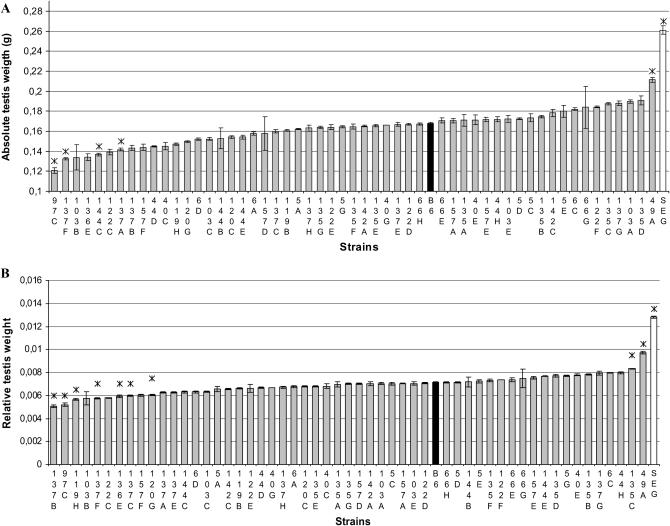
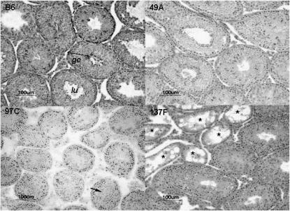
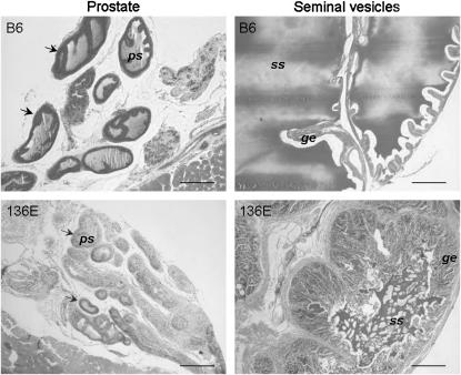
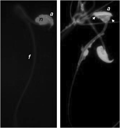
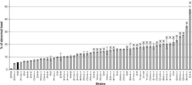
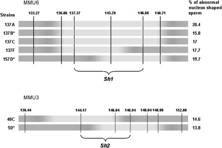
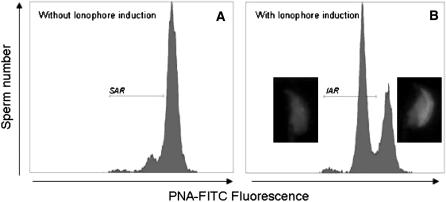
In mammals, male fertility is a quantitative feature determined by numerous genes. Until now, several wide chromosomal regions involved in fertility have been defined by genetic mapping approaches; unfortunately, the underlying genes are very difficult to identify. Here, 53 interspecific recombinant congenic mouse strains (IRCSs) bearing 1-2% SEG/Pas (Mus spretus) genomic fragments disseminated in a C57Bl/6J (Mus domesticus) background were used to systematically analyze male fertility parameters. One of the most prominent advantages of this model is the possibility of analyzing stable phenotypes in living animals. Here, we demonstrate the possibility in one-step fine mapping for several fertility traits. Focusing on strains harboring a unique spretus fragment, we could unambiguously localize two testis and one prostate weight-regulating QTL (Ltw1, Ltw2, and Lpw1), four QTL controlling the sperm nucleus shape (Sh1, Sh2, Sh3, and Sh4), and one QTL influencing sperm survival (Dss1). In several cases, the spretus DNA fragment was small enough to propose sound candidates. For instance, Spata1, Capza, and Tuba7 are very strong candidates for influencing the shape of the sperm head. Identifying new genes implied in mammalian fertility pathways is a necessary prerequisite for clarifying their molecular grounds and for proposing diagnostic tools for masculine infertilities.


Figures

F<sc>igure</sc> 1.—
Figure 1.—
Testis weight of parental and IRCS mice. The mean values (±SEM) of absolute (A) and relative (B) testis weight are presented in order of increasing values, with mean B6 and SEG testis weights shown in solid and open bars, respectively. Asterisks (*) denote strains that showed a significant difference in mean values compared to the B6 strain (P < 0.05).
F<sc>igure</sc> 2.—
Figure 2.—
Testis histology of three IRCS and B6 strains. (Top left) Seminiferous tubule sections (dotted lines) with normal spermatogenesis of B6 parental strain. These seminiferous tubules are used as a reference in the histological analysis of the IRCS (lu, lumen; ge, germinative epithelium). (Top right) Testis sections of the 49A strain, the IRCS with the heavier testes compared to B6; note the normal spermatogenesis and high cellular density in the seminiferous tubules, which have a diameter significantly superior to B6's diameter. (Bottom left) Testis section of the 97C strain. Most of the seminiferous tubules display an absence of lumen, but mature spermatozoa are detected (arrow); tubule diameters were significantly inferior to B6 ones. (Bottom right) Testis section of the 137F strain. Some tubules presented a SCO syndrome (asterisks); the remaining tubules display a normal spermatogenesis. Bar, 100 μm.
F<sc>igure</sc> 3.—
Figure 3.—
Histology of seminal vesicle and prostate of the 136E and B6 strains. When compared to B6 histology, 136E seminal vesicles presented reduced glandular lumens. Seminal secretions (ss), however, are visible, but in lesser quantity than in B6 glands. Glandular epithelium (ge) displays numerous convolutions, indicating a quiescent state of the gland. Prostate glands (arrows) of 136E present the same quiescent aspect as that of the seminal vesicles: no or very few prostate secretions (ps) can be observed in the gland lumens, compared to B6. Bar, 200 μm.
F<sc>igure</sc> 4.—
Figure 4.—
Morphology of mouse sperm from the cauda epididymis. Two fluorescent light microscope images of normal and abnormal (arrows) mouse spermatozoa stained with EH for the nucleus (n) and with FITC–PNA for the acrosome (a). (Right) Arrows point out the misshapenness of the head, which exhibited a normal-shaped acrosome (a); the flagellum (f) appears normal.
F<sc>igure</sc> 5.—
Figure 5.—
Frequency of sperm with an abnormal head in the IRCS. Mean values (±SEM) are presented in order of croissant abnormality frequency. B6 and SEG values are shown by solid bars. Asterisk (*) denotes 24 strains that showed a significant increase in mean values compared to the B6 strain.
F<sc>igure</sc> 6.—
Figure 6.—
Fine mapping of sperm hammerhead 1 and sperm hammerhead 2 QTL by an interstrain analysis. Marker positions are given in mega base pairs. Dark regions correspond to the B6 background and light regions to the SEG fragments in chromosomes 6 (MMU6) and 3 (MMU3) of different strains having comparable abnormal sperm-head frequency. Congenic strains are identified by an asterisk. Overlapping SEG fragments of the different strains makes it possible to map Shh1 QTL on a maximal 9-Mb fragment (137–146 Mb) on MMU6 and Shh2 QTL on a maximal 3 Mb of MMU3.
F<sc>igure</sc> 7.—
Figure 7.—
Flow cytometer analysis of acrosomal sperm status. (A) Flow cytometer acquisition in FITC spectra of non-ionophore-treated sperm: a population of spontaneous acrosome-reacted sperm exists under the SAR marker. (B) Flow cytometer acquisition in FITC spectra of ionophore-treated sperm: a population of induced acrosome-reacted sperm appears under the IAR marker.

Similar articles

Cited by

References

    1. Acampora, D., S. Mazan, F. Tuorto, V. Avantaggiato, J. J. Tremblay et al., 1998. Transient dwarfism and hypogonadism in mice lacking Otx1 reveal prepubescent stage-specific control of pituitary levels of GH, FSH and LH. Development 125: 1229–1239. - PubMed
    1. Affara, N. A., and M. J. Mitchell, 2000. The role of human and mouse Y chromosome genes in male infertility. J. Endocrinol. Invest. 23: 630–645. - PubMed
    1. Avner, P., 1998. Complex traits and polygenic inheritance in the mouse. Methods 14: 191–198. - PubMed
    1. Bolor, H., N. Wakasugi, W. D. Zhao and A. Ishikawa, 2006. Detection of quantitative trait loci causing abnormal spermatogenesis and reduced testis weight in the small testis (Smt) mutant mouse. Exp. Anim. 55: 97–108. - PubMed
    1. Carlson, A. E., R. E. Westenbroek, T. Quill, D. Ren, D. E. Clapham et al., 2003. CatSper1 required for evoked Ca2+ entry and control of flagellar function in sperm. Proc. Natl. Acad. Sci. USA 100: 14864–14868. - PMC - PubMed

Publication types