. 2007 Jul;176(3):1483-90.
doi: 10.1534/genetics.107.071134. Epub 2007 May 4.

Meiotic parthenogenesis in a root-knot nematode results in rapid genomic homozygosity

Affiliations

Meiotic parthenogenesis in a root-knot nematode results in rapid genomic homozygosity

Qingli L Liu et al. Genetics. 2007 Jul.

Abstract

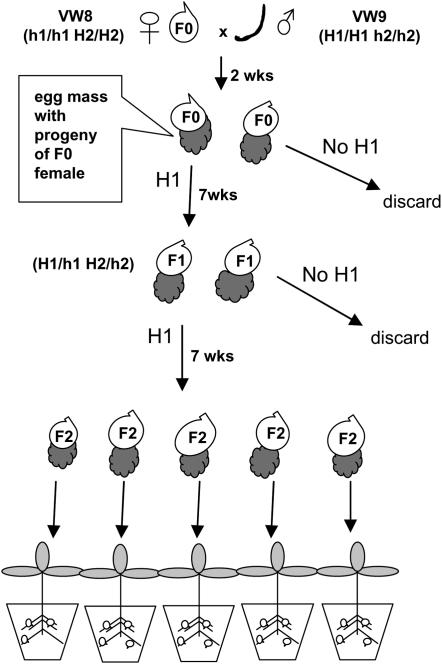
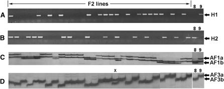
Many isolates of the plant-parasitic nematode Meloidogyne hapla reproduce by facultative meiotic parthenogenesis. Sexual crosses can occur, but, in the absence of males, the diploid state appears to be restored by reuniting sister chromosomes of a single meiosis. We have crossed inbred strains of M. hapla that differ in DNA markers and produced hybrids and F(2) lines. Here we show that heterozygous M. hapla females, upon parthenogenetic reproduction, produce progeny that segregate 1:1 for the presence or absence of dominant DNA markers, as would be expected if sister chromosomes are rejoined, rather than the 3:1 ratio typical of a Mendelian cross. Codominant markers also segregate 1:1 and heterozygotes are present at low frequency (<3%). Segregation patterns and recombinant analysis indicate that a homozygous condition is prevalent for markers flanking recombination events, suggesting that recombination occurs preferentially as four-strand exchanges at similar locations between both pairs of non-sister chromatids. With this mechanism, meiotic parthenogenesis would be expected to result in rapid genomic homozygosity. This type of high negative crossover interference coupled with positive chromatid interference has not been observed in fungal or other animal systems in which it is possible to examine the sister products of a single meiosis and may indicate that meiotic recombination in this nematode has novel features.


Figures

F<sc>igure</sc> 1.—
Figure 1.—
Strategy for producing F2 lines of M. hapla. A culture with young females (F0) of strain VW8 (which lacks PCR marker H1) is inoculated with males (curved line) of strain VW9 (which carries marker H1). After 2 weeks, egg masses are collected from VW8 F0 females and tested for the presence of marker H1. Juveniles from egg masses with marker H1 are inoculated onto plants and allowed to develop parthenogenetically into F1 females. Egg masses from F1 females are tested for the presence of marker H1. Eggs from F2 egg masses are inoculated onto individual plants. This figure is adapted from Williamson and Liu (2006).
F<sc>igure</sc> 2.—
Figure 2.—
DNA marker segregation in F2 lines from hybrid females of M. hapla. For each marker, phenotypes of parental lines VW8 and VW9 are shown at the right. Lanes marked “F2 lines” show DNA from individual lines amplified with the PCR primers H1 (A) or H2 (B). Segregation pattern of allelic AFLP markers AF1a and AF1b in F2 lines is shown in C. The monomorphic band between AF1a and AF1b is a useful control for PCR amplification. (D) Segregation pattern of allelic AFLP markers AF3a and AF3b in F2 lines. The one heterozygous line is designated with an “x.”
F<sc>igure</sc> 3.—
Figure 3.—
Cytology of meiotic maturation in M. hapla. (A–F) Successive images from the same gonad stained with Hoescht 33258 that represent progression from oocytes just posterior to the spermatheca. The gonad was from a culture of strain VW9 infected with a low nematode innoculum. No sperm were seen in the spermatheca of this gonad. (A) Multiple oocytes arrested in metaphase I. (B) The last metaphase I oocyte (labeled M) is followed by an oocyte that appears to be in anaphase II. The following oocyte (C) appears to be arrested in telophase II with two pronuclei and a condensed polar body or polar nucleus. As the oocytes move toward the vagina, the two pronuclei are seen progressively closer and merge together (D–F). The polar nucleus or polar body from meiosis I is indicated by an arrowhead. In E, the polar body appears to be extruded from the cell and no polar body is apparent in F. Bar in A–F, 0.02 mm.
F<sc>igure</sc> 4.—
Figure 4.—
Model for recombination and segregation in M. hapla. (A) Linkage group with four codominant AFLP markers. Numbers to the left represent the percentage of recombinant chromosomes between markers based on the data in Table 2. (B) Model to explain double recombinants between markers AF22 and AF24. (1) A four-strand double exchange occurs between markers AF22 and AF24. (2) Open arrows show the direction of movement of chromosomes during anaphase I and telophase II. (3) One set of sister chromosomes forms polar body I and the other set undergoes a second meiotic division arresting in telophase II. (4) In the absence of sperm nucleus fusion, the sister chromosomes are rejoined in a single pronucleus homozygous for all markers.

References

    1. Albertson, D. G., A. M. Rose and A. M. Villeneuve, 1997. Chromosome organization, mitosis, and meiosis, pp. 47–78 in C. elegans II, edited by D. L. Riddle, T. Blumenthal, B. J. Meyer and J. R. Priess. Cold Spring Harbor Laboratory Press, Cold Spring Harbor, NY. - PubMed
    1. Asher, J. H., 1970. Pathenogenesis and genetic variability. II. One-locus models for various diploid populations. Genetics 66: 369–391. - PMC - PubMed
    1. Atibalentja, N., S. Bekal, L. L. Domier, T. L. Niblack, G. R. Noel et al., 2005. A genetic linkage map of the soybean cyst nematode Heterodera glycines. Mol. Gen. Genomics 273: 273–281. - PubMed
    1. Barker, K. R., 1985. Nematode extraction and bioassays, pp. 19–35 in An Advanced Treatise on Meloidogyne, Vol II, edited by K. R. Barker, C. C. Carter and J. N. Sasser. North Carolina University Graphics, Raleigh, NC.
    1. Barker, K. R., and S. R. Koenning, 1998. Developing sustainable systems for nematode management. Annu. Rev. Phytopathol. 36: 165–205. - PubMed

Publication types