. 2007 Nov;177(3):1527-37.
doi: 10.1534/genetics.107.070771. Epub 2007 May 4.

Fine-tuning of translation termination efficiency in Saccharomyces cerevisiae involves two factors in close proximity to the exit tunnel of the ribosome

Affiliations

Fine-tuning of translation termination efficiency in Saccharomyces cerevisiae involves two factors in close proximity to the exit tunnel of the ribosome

Isabelle Hatin et al. Genetics. 2007 Nov.

Abstract

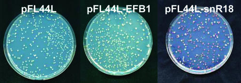
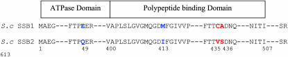
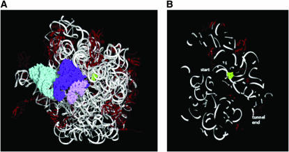
In eukaryotes, release factors 1 and 3 (eRF1 and eRF3) are recruited to promote translation termination when a stop codon on the mRNA enters at the ribosomal A-site. However, their overexpression increases termination efficiency only moderately, suggesting that other factors might be involved in the termination process. To determine such unknown components, we performed a genetic screen in Saccharomyces cerevisiae that identified genes increasing termination efficiency when overexpressed. For this purpose, we constructed a dedicated reporter strain in which a leaky stop codon is inserted into the chromosomal copy of the ade2 gene. Twenty-five antisuppressor candidates were identified and characterized for their impact on readthrough. Among them, SSB1 and snR18, two factors close to the exit tunnel of the ribosome, directed the strongest antisuppression effects when overexpressed, showing that they may be involved in fine-tuning of the translation termination level.


Figures

F<sc>igure</sc> 1.—
Figure 1.—
FS1 modified strain with ADE2 locus reporter was transformed by pFL44L vector empty, pFL44L-eEF1Bα, or pFL44L-snR18 and spread on CSM minimal medium. The color intensity was checked after incubation for 5 days at 30°.
F<sc>igure</sc> 2.—
Figure 2.—
FS1 and 74D694 [psi−] or [psi+] strains were cotransformed with pAC-TMG vector bearing a UAG stop codon and SSB1 expressed either from centromeric (pCMSSB1) or multicopy (pYESSB1) vector. The readthrough level was expressed as the percentage of readthrough decrease referred to the empty vector.
F<sc>igure</sc> 3.—
Figure 3.—
The readthrough level in MT557/3b strain was quantified by the dual reporter system pAC with the three stop codon targets TAA, TAG, or TGA in the presence or absence of overexpressed Sup45p, Ssb1p, or Ssb2p.
F<sc>igure</sc> 4.—
Figure 4.—
The MT557/3b strain transformed or not transformed by mutated overexpressed proteins Sup45p, Ssb2p, Ssb1p, or ssb1p was incubated at 30° and 37° for 3 days.
F<sc>igure</sc> 5.—
Figure 5.—
Four amino acids differ between S. cerevisiae SSB1 and SSB2. These proteins show an ATPase domain from amino acid 1 to 400 and a polypeptide-binding domain from amino acid 401 to 507.
F<sc>igure</sc> 6.—
Figure 6.—
snR18 guides modification to sites in the wall of the polypeptide exit tunnel. (A) The large ribosomal subunit viewed down the polypeptide exit tunnel, with the start of the tunnel in the front of the image and the end, where the nascent polypeptide emerges, in the back. Proteins (red) and RNA as ribbon representation. The nucleotides targeted for 2′-O-methylation due to complementarity between the rRNA and snR18 are highlighted by showing their van der Waals radii (green). Starting in the canonical “crown view,” the subunit (of Thermus thermophilus; pdb code 2j01) has been rotated forward slightly around the horizontal axis and slightly counterclockwise around the vertical axis. The fragment of A-site tRNA visible in the complex is pink; a complete P-site tRNA (purple) and E-site tRNA (cyan) is observed. Importantly, the targeted sites occur in the conserved core of the large subunit (Gerbi 1996), validating examining structures from other species. (B) Cross section of the large ribosomal subunit (a 13.2 Å thick slab) as viewed from the side to feature the polypeptide exit tunnel. Details are as in A, except the tRNAs are not shown. This view is obtained by rotation of 90° about the vertical axis from the crown view and rotating the subunit backward slightly around the horizontal axis.

Similar articles

Cited by

References

    1. Alkalaeva, E. Z., A. V. Pisarev, L. Y. Frolova, L. L. Kisselev and T. V. Pestova, 2006. In vitro reconstitution of eukaryotic translation reveals cooperativity between release factors eRF1 and eRF3. Cell 125: 1125–1136. - PubMed
    1. Andjelkovic, N., S. Zolnierowicz, C. Van Hoof, J. Goris and B. A. Hemmings, 1996. The catalytic subunit of protein phosphatase 2A associates with the translation termination factor eRF1. EMBO J. 15: 7156–7167. - PMC - PubMed
    1. Bailleul, P. A., G. P. Newnam, J. N. Steenbergen and Y. O. Chernoff, 1999. Genetic study of interactions between the cytoskeletal assembly protein sla1 and prion-forming domain of the release factor Sup35 (eRF3) in Saccharomyces cerevisiae. Genetics 153: 81–94. - PMC - PubMed
    1. Barton-Davis, E. R., L. Cordier, D. I. Shoturma, S. E. Leland and H. L. Sweeney, 1999. Aminoglycoside antibiotics restore dystrophin function to skeletal muscles of mdx mice. J. Clin. Invest. 104: 375–381. - PMC - PubMed
    1. Beckmann, R. P., L. E. Mizzen and W. J. Welch, 1990. Interaction of Hsp 70 with newly synthesized proteins: implications for protein folding and assembly. Science 248: 850–854. - PubMed

Publication types

MeSH terms