. 2007 May 8;104(19):8178-83.
doi: 10.1073/pnas.0700865104. Epub 2007 May 2.

Conserved domain structure of pentatricopeptide repeat proteins involved in chloroplast RNA editing

Affiliations

Conserved domain structure of pentatricopeptide repeat proteins involved in chloroplast RNA editing

Kenji Okuda et al. Proc Natl Acad Sci U S A. .

Abstract

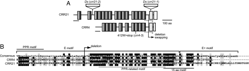
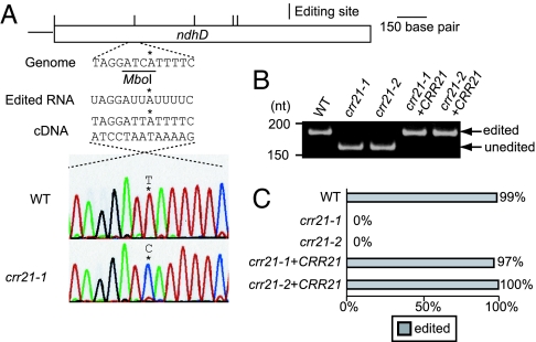
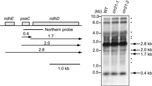
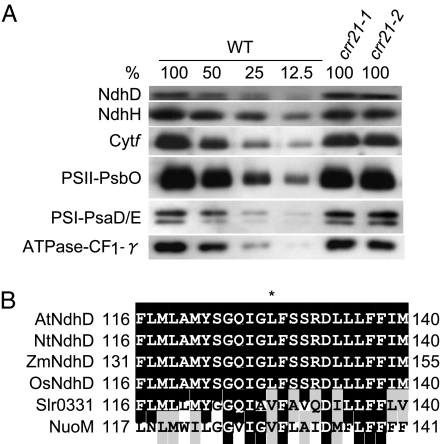
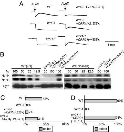
The pentatricopeptide repeat (PPR) proteins form one of the largest families in higher plants and are believed to be involved in the posttranscriptional processes of gene expression in plant organelles. It has been shown by using a genetic approach focusing on NAD(P)H dehydrogenase (NDH) activity that a PPR protein CRR4 is essential for a specific RNA editing event in chloroplasts. Here, we discovered Arabidopsis crr21 mutants that are specifically impaired in the RNA editing of the site 2 of ndhD (ndhD-2), which encodes a subunit of the NDH complex. The CRR21 gene encodes a member of the PPR protein family. The RNA editing of ndhD-2 converts the Ser-128 of NdhD to leucine. In crr21, the activity of the NDH complex is specifically impaired, suggesting that the Ser128Leu change has important consequences for the function of the NDH complex. Both CRR21 and CRR4 belong to the E+ subgroup in the PLS subfamily that is characterized by the presence of a conserved C-terminal region (the E/E+ domain). This E/E+ domain is highly conserved and exchangeable between CRR21 and CRR4, although it is not essential for the RNA binding. Our results suggest that the E/E+ domain has a common function in RNA editing rather than of recognizing specific RNA sequences.


Conflict of interest statement

The authors declare no conflict of interest.

Figures

Fig. 1.
Fig. 1.
Monitoring of NDH activity by using chlorophyll fluorescence analysis. The curve shows a typical trace of chlorophyll fluorescence in the wild type (WT). Leaves were exposed to AL (50 μmol of photons m−2 s−1) for 5 min. AL was turned off and the subsequent change in chlorophyll fluorescence level was monitored. Insets are magnified traces from the boxed area. The fluorescence levels were normalized by Fm levels. ML, measuring light; SP, saturating pulse of white light; crr21+CRR21, crr21 complemented by introduction of the wild-type genomic CRR21.
Fig. 2.
Fig. 2.
Predicted motif structure of CRR21. (A) Predicted motif structure of CRR21 as compared with that of CRR4. PPR motifs are depicted as shaded black boxes. The E motif is shown as a dotted line. The PPR-related motif in the E motif and the E+ motif are depicted as gray and white boxes, respectively. The putative transit peptide to plastids is underlined. The 15-amino acid motif is shown by an asterisk. Sites of Ds insertion and the position of the crr4–3 mutation (nonsense) are indicated. Positions of the deletion and domain swapping are indicated. (B) Partial sequence alignment of CRR21 and CRR4. Alignment was performed by using the ClustalW program. The consensus sequence of the E and E+ motifs according to bioinformatics analysis by Lurin et al. (5) is shown at the top of the sequences. The best-conserved residues are in capital letters. Amino acids that are fully conserved or substitutive are respectively shaded black and gray. The PPR-related motif in the E motif and the 15-amino acid motif are respectively underlined by a dotted line and a solid line. The last PPR motifs of CRR4 and CRR21 are indicated. The deletion of the E/E+ domain of CRR4 in the crr4–3+CRR4 (−E/E+) construct is specified.
Fig. 3.
Fig. 3.
Analysis of RNA editing of ndhD-2. (A) Direct sequencing of RT-PCR products containing the ndhD-2 editing site. Positions of RNA editing are indicated. The restriction enzyme MboI specifically cleaves cDNA derived from unedited molecules. The ndhD-2 site is shown by an asterisk. (B) Semiquantitative analysis of the extent of RNA editing. RT-PCR products were digested with MboI. Fragments originating from edited and unedited RNA molecules are indicated. (C) The editing efficiency of ndhD-2 was analyzed as described in Materials and Methods. Ratio of clones originated from edited RNA molecules is indicated by gray bars. crr21+CRR21, crr21 complemented by introduction of the wild-type genomic CRR21.
Fig. 4.
Fig. 4.
Transcript pattern of psaC-ndhD operon. Total RNA (5 μg) isolated from leaves of 3-week-old wild type (Nössen), crr21–1, and crr21–2 was analyzed by RNA gel blot hybridization. The positions of RNA size markers are indicated on the left. The positions and sizes of RNA detected by RNA gel blot hybridization indicated by asterisks and arrows with letters on the right. Transcript map was deduced from this and the previous report (41).
Fig. 5.
Fig. 5.
Protein blot analysis of the NDH complex and the major photosynthetic complexes. (A) Immunodetection of NDH subunits, NdhD and NdhH; a subunit of the cytb6f complex, Cytf; subunits of the photosystem I, PsaE/D; a subunit of the photosystem II, PsbO; and γ-subunit of the chloroplast F0F1-ATPase, CF1-γ. Proteins were extracted from the thylakoid membrane fractions. Lanes were loaded with protein samples corresponding to 0.5 μg chlorophyll for Cytf, PsaD/E, PsbO, and CF1-γ, 5 μg chlorophyll for NdhH, and 10 μg chlorophyll for NdhD (100%) and the series of dilutions indicated. (B) Partial sequence alignment of NdhD containing the editing site. Arabidopsis NdhD protein was aligned with their homologous proteins. Alignment was performed by using the ClustalW program. Sequences (GenBank accession numbers) shown here are as follows: Arabidopsis thaliana (AtNdhD, BAA84437), Nicotiana tabacum (NtNdhD, CAA77432), Zea mays (ZmNdhD, CAA31558), Oryza sativa (OsNdhD, NP_039444), Synechocystis sp. PCC 6803 (Slr0331, NP_441967), and Escherichia coli K12 (NuoM, NP_416780). Amino acids that are fully conserved or substitutive are shaded black and gray, respectively. The asterisk indicates the position of an edited codon. The Ser-128 of AtNdhD and NtNdhD is converted to leucine by RNA editing of ndhD-2. Numbers indicate amino acid positions in protein.
Fig. 6.
Fig. 6.
Effects of the deletion and conversion of the E/E+ domain in CRR4. (A) Analysis of the transient increase in chlorophyll fluorescence after turning off AL. Leaves were exposed to AL (50 μmol of photons m−2 s−1) for 5 min. AL was turned off, and the subsequent change in chlorophyll fluorescence level was monitored. The fluorescence levels were normalized by Fm levels. (B) Protein blot analysis of the NDH complex. Immunodetection of NDH subunits, NdhD and NdhH, and a subunit of the cytb6f complex, Cytf. Proteins were extracted from the thylakoid membrane fractions. Lanes were loaded with protein samples corresponding to 0.5 μg of chlorophyll for Cytf, 5 μg of chlorophyll for NdhH, and 10 μg of chlorophyll for NdhD (100%) and the series of dilutions indicated. (C) Analysis of the extent of RNA editing in the ndhD initiation codon. The editing efficiency of the ndhD-1 was analyzed as described in Materials and Methods. Ratio of clones originated from edited RNA molecule is indicated by gray bars. (D) Analysis of the extent of RNA editing in the ndhD-2 site. Ratio of clones originated from edited RNA molecule is indicated by gray bars. crr4–3+CRR4 (−E/E+), crr4–3 transformed with the CRR4 truncated in the E/E+ domain; crr4–3+CRR4 (+21E/E+), crr4–3 transformed with CRR4, in which the E/E+ domain was replaced by that of CRR21; crr21–1+CRR21(+4E/E+), crr21–1 transformed with CRR21, in which the E/E+ domain was replaced by that of CRR4.

Similar articles

Cited by

References

    1. Mullet JE. Annu Rev Plant Physiol Plant Mol Biol. 1988;39:475–502. - PubMed
    1. Hedtke B, Börner T, Weihe A. Science. 1997;277:809–811. - PubMed
    1. Deng XW, Gruissem W. Cell. 1987;49:379–387. - PubMed
    1. Barkan A, Goldshumidt-Clermont M. Biochimie. 2000;82:559–572. - PubMed
    1. Lurin C, Andres C, Aubourg S, Bellaoui M, Bitton F, Bruyere C, Caboche M, Debast C, Gualberto J, Hoffmann B, et al. Plant Cell. 2004;16:2089–2103. - PMC - PubMed

Publication types