. 2007 May 15;104(20):8340-5.
doi: 10.1073/pnas.0700076104. Epub 2007 May 1.

Decondensing the protamine domain for transcription

Affiliations

Decondensing the protamine domain for transcription

Rui Pires Martins et al. Proc Natl Acad Sci U S A. .

Abstract

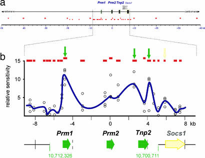
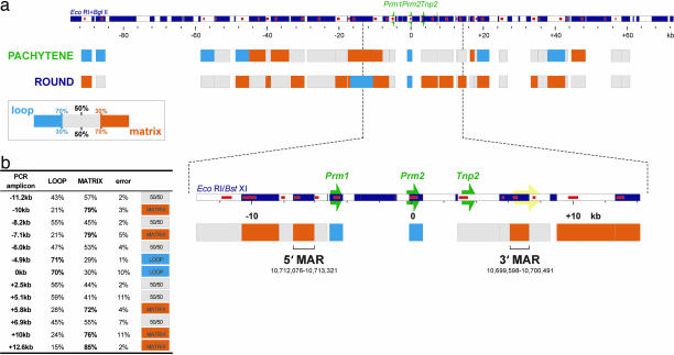
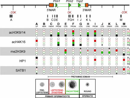
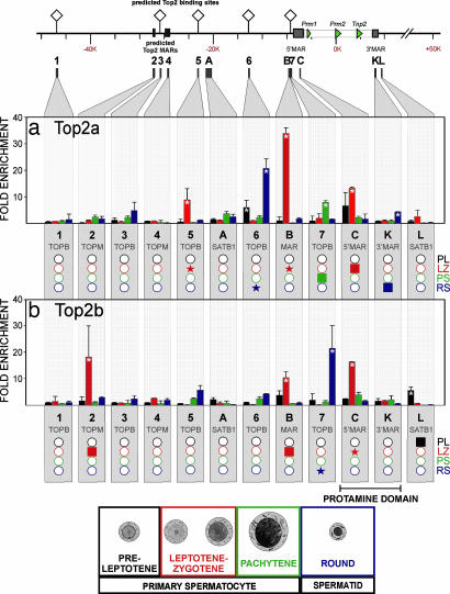
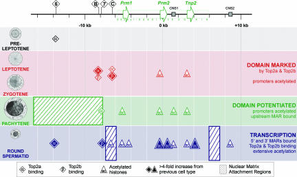
Potentiation is the transition from higher-order, transcriptionally silent chromatin to a less condensed state requisite to accommodating the molecular elements required for transcription. To examine the underlying mechanism of potentiation an approximately 13.7-kb mouse protamine domain of increased nuclease sensitivity flanked by 5' and 3' nuclear matrix attachment regions was defined. The potentiated DNase I-sensitive region is formed at the pachytene spermatocyte stage with the recruitment to the nuclear matrix of a large approximately 9.6-kb region just upstream of the domain. Attachment is then specified in the transcribing round spermatid, recapitulating the organization of the human cluster. In comparison to other modifiers that have no effect, i.e., histone methylation, HP1, and SATB1, topoisomerase engages nuclear matrix binding as minor marks of histone acetylation appear. Reorganization is marked by specific sites of topoisomerase II activity that are initially detected in leptotene-zygotene spermatocytes just preceding the formation of the DNase I-sensitive domain. This has provided a likely model of the events initiating potentiation, i.e., the opening of a chromatin domain.


Conflict of interest statement

The authors declare no conflict of interest.

Figures

Fig. 1.
Fig. 1.
Definition of the DNase I-sensitive region encompassing the mouse protamine gene cluster. (a) DNase I sensitivity was initially assayed in round spermatid nuclei at 12 sites throughout a 90-kb region of mouse chromosome 16qA1 encompassing the protamine cluster using real-time PCR (upper row of small red boxes; see supporting information (SI) Table 1 for oligonucleotide sequences). A second, higher-resolution analysis of a subregion included an additional 21 amplicons (lower row of small red boxes). (b) Data from 21 amplicons (red boxes) immediately encompassing the gene cluster from four independent experiments is shown as a function of genomic position (open circles). A running average of the median of the four points is plotted in blue, identifying an ≈11.6-kb DNase I-sensitive domain encompassing coordinates 10,700,711–10,712,326 (National Center for Biotechnology Information public mouse genome assembly Build 36.1). DNase I-hypersensitive sites within the region assayed are shown as peaks and are marked by arrows.
Fig. 2.
Fig. 2.
Nuclear matrix associations encompassing the mouse protamine domain. (a) Long-range nuclear matrix association of an ≈142-kb region encompassing the mouse protamine domain was assayed in pachytene primary spermatocytes (potentiated domain) and round spermatids (potentiated and transcribed domain). Nuclei were treated with 2 M NaCl, then restricted with EcoRI and BglII to release nonmatrix-associated, i.e., loop DNA. DNA was fractionated and then purified, and degree of nuclear matrix association for 32 restriction fragments was assayed by real-time PCR. Nuclear matrix binding potentials were calculated for the interrogated regions. Fragments that were highly enriched in the nuclear matrix-associated fraction (≥70% matrix/≤30% loop), termed matrix-attached, are shown in orange. Fragments that were highly enriched in the loop fraction (≥70% loop/≤30% matrix) are shown in cyan. All other regions, termed matrix-associated, are shown as gray boxes. A large ≈9.6-kb MAR upstream of the domain binds in pachytene spermatocytes. A considerable increase in the number of MARs was observed in round spermatids. (b) EcoRI and BstXI were used to resolve the round spermatid nuclear MARs. Nuclear matrix binding potentials for the 13 restriction fragments interrogated about the protamine domain are shown in the table (error displayed is one standard deviation of the mean for two independent experiments). Two restriction fragments, one upstream of Prm1 (coordinates 10,712,076–10,713,321) and another downstream of Tnp2 (10,699,598–10,700,491) demarcated the 5′ MAR and 3′ MAR, respectively. Genes of the protamine domain are indicated by green arrows, the neighboring Socs1 gene is indicated in yellow, restriction fragments are shown as alternating white and dark blue boxes, and PCR amplicons are indicated as red boxes within restriction maps.
Fig. 3.
Fig. 3.
Epigenetic modification of the mouse protamine domain during spermatogenesis. Histone acetylation (acH3K9/14 and acH4K16), association of HP1 and SATB1, and histone methylation (me2H3K9) of the protamine domain were assessed by ChIP at 13 sites (A–M) in preleptotene (black; PL), leptotene–zygotene (red; LZ), and pachytene (green; PS) primary spermatocytes, as well as round spermatids (blue; RS). These included gene promoters (PROM: regions D, F, and I), coding sequences (CDS: regions E, G, and J), experimentally confirmed MARs (regions B, C, and K), CNS (regions H, and L). Regions A and M are upstream and downstream of the domain, respectively. Shaded boxes are those regions yielding a signal that was significantly greater than the preceding cell type in the spermatogenic pathway. Shaded stars represent a >4-fold increase compared with the preceding cell type. Open circles indicate regions with no significant association.
Fig. 4.
Fig. 4.
Association of topoisomerase II with the mouse protamine domain during spermatogenesis. The binding of topoisomerase II α (Top2a) and β (Top2b) to the mouse protamine domain was assessed by ChIP at 12 sites (1–7, regions A, B, K, and L) in preleptotene, leptotene–zygotene, and pachytene primary spermatocytes as well as round spermatids. Sites included predicted Top2 binding sites (TOPB: 1, 3, and 5–7), predicted Top2 MARs (TOPM: 2 and 4), and experimentally confirmed MARs (regions B, C, and K), as well as sites flanking the domain that showed association with SATB1 in round spermatids (regions A and L). The region encompassing the 5′ MAR exhibits differential Top2a (a) and Top2b (b) associations throughout spermatogenesis. Values presented are means, and error bars represent the 95% confidence intervals. Asterisks denote a significant increase (P < 0.05) in that modification compared with the preceding cell types in the spermatogenic pathway. Below each panel, the data are summarized with shaded boxes representing those regions yielding a signal that was significantly greater than the preceding cell type in the spermatogenic pathway. Shaded stars represent a >4-fold increase compared with the preceding cell type. Open circles indicate regions with no significant association.
Fig. 5.
Fig. 5.
The molecular events that precede transcription of the protamine domain. Before acquiring a potentiated conformation in the pachytene spermatocyte, the mouse protamine domain is marked at upstream sites B and C by Top2a and Top2b in leptotene and zygotene spermatocytes. The promoters of Prm2 and Tnp2 are marked by histone (H3K9/14) acetylation. Coincident with the potentiation of the domain, a large region immediately upstream of the Top2 cleavage sites of the gene cluster is recruited to the nuclear matrix. The promoters of all three genes and CNS1 within the domain are acetylated. A single site (site 7) of Top2a activity was detected toward the proximal end of this ≈9.6-kb MAR in pachytene spermatocytes. The genes of the protamine domain are transcribed postmeiotically in round spermatids. At this time, two MARs that flank the domain bind the nuclear matrix in conjunction with extensive histone acetylation throughout the domain and a single upstream CNS2. Significant induction of Top2a site 6 and Top2b site 7 activity were detected upstream of the domain. Top2 functions in concert with other nuclear matrix factors to mark this multigenic domain for potentiation and eventual transcription.

Similar articles

Cited by

References

    1. Martins RP, Krawetz SA. Gene Ther Mol Biol. 2005;9:229–246. - PMC - PubMed
    1. Choudhary SK, Wykes SM, Kramer JA, Mohamed AN, Koppitch F, Nelson JE, Krawetz SA. J Biol Chem. 1995;270:8755–8762. - PubMed
    1. Chambeyron S, Da Silva NR, Lawson KA, Bickmore WA. Development (Cambridge, UK) 2005;132:2215–2223. - PubMed
    1. Chambeyron S, Bickmore WA. Genes Dev. 2004;18:1119–1130. - PMC - PubMed
    1. Keene MA, Corces V, Lowenhaupt K, Elgin SCR. Eur J Cell Biol. 1980;22:95–95.

Publication types

LinkOut - more resources