. 2007 Oct;122(2):157-66.
doi: 10.1111/j.1365-2567.2007.02621.x. Epub 2007 May 2.

Expression of 5-lipoxygenase (5-LOX) in T lymphocytes

Affiliations

Expression of 5-lipoxygenase (5-LOX) in T lymphocytes

Jeanne M Cook-Moreau et al. Immunology. 2007 Oct.

Abstract

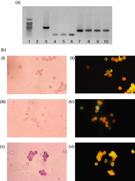
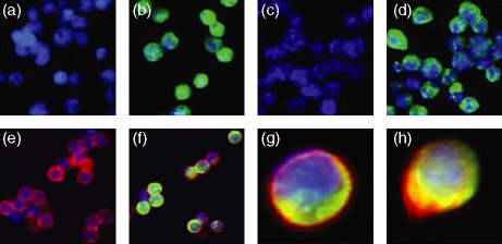
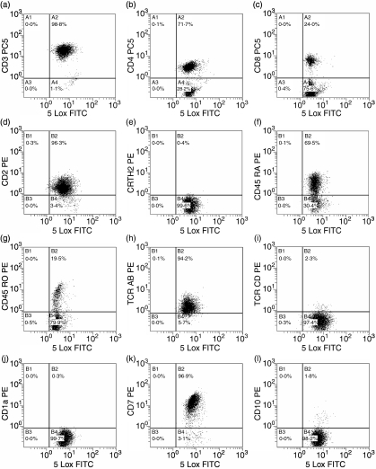
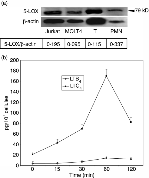
5-Lipoxygenase (5-LOX) is the key enzyme responsible for the synthesis of the biologically active leukotrienes. Its presence has been reported in cells of the myeloid lineage and B lymphocytes but has not been formally defined in T lymphocytes. In this study, we provide evidence for 5-LOX expression on both transcriptional and translational levels in highly purified peripheral blood T cells as well as in human T lymphoblastoid cell lines (MOLT4 and Jurkat). Messenger RNA (mRNA) of 5-LOX was amplified by conventional reverse transcription-polymerase chain reaction (RT-PCR; MOLT4 and Jurkat cells) and by in situ RT-PCR (T lymphocytes). 5-LOX protein expression was confirmed by Western blot and immunofluorescence studies. 5-LOX was present primarily in the cytoplasm with some nuclear localization and was translocated to the nuclear periphery after culture in a mitosis-supporting medium. Fluorescence-activated cell sorter analysis of different T-lymphocyte populations, including CD4, CD8, CD45RO, CD45RA, T helper type 2, and T-cell receptor-alphabeta and -gammadelta expressing cells, did not identify a differential distribution of the enzyme. Purified peripheral blood T lymphocytes were incapable of synthesizing leukotrienes in the absence of exogenous arachidonic acid. Jurkat cells produced leukotriene C(4) and a small amount of leukotriene B(4) in response to CD3-CD28 cross-linking. This synthesis was abolished by two inhibitors of leukotriene synthesis, MK-886 and AA-861. The presence of 5-LOX in T lymphocytes but the absence of endogenous lipoxygenase metabolite production compared to Jurkat cells may constitute a fundamental difference between resting peripheral lymphocytes and leukaemic cells.


Figures

Figure 1
Figure 1
Expression of 5-LOX mRNA by MOLT4 and Jurkat T cell lines as well as peripheral blood T cells. (a) Amplification of 5-LOX mRNA by RT-PCR (35 cycles). Lane 1, molecular weight ladder; lane 2, negative control – cDNA from chloramphenicol acetyl transferase mRNA with 5-LOX primers; lane 3, positive control – same cDNA with corresponding primers; lane 4, Jurkat mRNA run without reverse transcriptase; lane 5, MOLT4 mRNA run without reverse transcriptase; lane 6, T-lymphocyte mRNA run without reverse transcriptase; lane 7, 5-LOX cDNA-positive control; lane 8, Jurkat cells; lane 9, MOLT4 cells; lane 10, purified peripheral T lymphocytes. (b) Amplification of 5-LOX mRNA by RT-PCR in situ. Panel i, negative control: peripheral blood T lymphocytes submitted to RT-PCR in situ without reverse transcriptase; panel ii, same field visualized by fluorescence microscopy after incubation with anti-CD3 antibodies; panel iii, negative control: peripheral blood T lymphocytes submitted to RT-PCR in situ with primers specific for chloramphenicol acetyl transferase; panel iv, same field visualized by fluorescence microscopy after incubation with anti-CD3 antibodies, panel v, peripheral blood T lymphocytes submitted to RT-PCR in situ with specific 5-LOX primers; panel vi, same field visualized by fluorescence microscopy after incubation with anti-CD3 antibodies. Magnification 65×.
Figure 2
Figure 2
5-LOX expression in Jurkat and MOLT4 cell lines and peripheral blood T lymphocytes by indirect immunofluorescence. (a and c) Jurkat and MOLT4 cells, respectively, incubated with a negative isotypic control serum; (b and d) Jurkat and MOLT4 cells, respectively, incubated with anti-5-LOX monoclonal antibody; (e and f) peripheral blood T lymphocytes incubated with either a negative isotypic control serum (e) or anti-5-LOX monoclonal antibody (f) followed by staining with anti-CD3 monoclonal antibodies (magnification × 40); (g) peripheral blood T lymphocytes stained with anti-5-LOX and anti-CD3 monoclonal antibodies and (h) after 3 days incubation in Chang MF medium and stained with anti-5-LOX and anti-CD3 monoclonal antibodies (arrow indicates localization of 5-LOX at nuclear periphery).
Figure 3
Figure 3
Analysis of peripheral blood T lymphocyte populations by flow cytometry. The acquisition panels represent T cells gated on the CD19-negative, CD45-positive population. Cells were incubated with anti-5-LOX monoclonal antibody and F(ab)′2 goat anti-mouse fluorescein isothiocyanate (FITC) followed by staining with anti-CD3 (a), CD4 (b), CD8 (c), CD2 (d), CRTH2 (e), CD45RA (f), CD45RO (g), TCR-αβ (h), TCR-γδ (i), CD1a (j), CD7 (k) and CD10 (l) antibodies. The x-axis represents 5-LOX FITC and the y-axis represents the antibodies defining the different subpopulations. Results are from one experiment and are representative of five.
Figure 4
Figure 4
5-LOX protein expression and metabolite production. (a) Western blot of 5-LOX in MOLT4 and Jurkat T-cell lines and peripheral blood T lymphocytes. Cells were lysed and the cytosolic fraction was retrieved by ultracentrifugation. Equal amounts of protein (25 μg) were subjected to SDS–PAGE on 4–15% gradient gels, then transferred and immunoblotted using anti-5-LOX monoclonal antibody. Lane 1, molecular weight standard; lane 2, Jurkat cells; lane 3, MOLT4 cells; lane 4, peripheral blood T cells; lane 5, neutrophils. (b) Synthesis of LTB4 and LTC4 by Jurkat cells. Cells were activated by CD2–CD3–CD28 crosslinking. Supernatants were harvested at different times and production of LTB4 and LTC4 was quantified by EIA.

Similar articles

Cited by

References

    1. Funk CD. Prostaglandins and leukotrienes: advances in eicosanoid biology. Science. 2001;294:1871–5. - PubMed
    1. Ruegg C, Dormond O, Mariotti A. Endothelial cell integrins and COX-2: mediators and therapeutic targets of tumor angiogenesis. Biochim Biophys Acta. 2004;1654:51–67. - PubMed
    1. Goodwin JS, Ceuppens J. Regulation of the immune response by prostaglandins. J Clin Immunol. 1983;3:295–315. - PubMed
    1. Stjernschantz J. The leukotrienes. Med Biol. 1984;62:215–30. - PubMed
    1. Rouzer CA, Kargman S. Translocation of 5-lipoxygenase to the membrane in human leukocytes challenged with ionophore A23187. J Biol Chem. 1988;263:10980–8. - PubMed

MeSH terms