Viscoelastic measurements of vocal folds using the linear skin rheometer
- PMID: 17485196
- PMCID: PMC7994085
- DOI: 10.1016/j.jvoice.2007.01.002
Viscoelastic measurements of vocal folds using the linear skin rheometer
Abstract
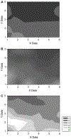
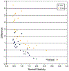
As the number of interventions for vocal fold scar grows and with the advancement of mathematical modeling, greater accuracy and precision in the measurement of vocal fold pliability will become essential. Although indirect pliability measures have been used successfully, direct measurement of tissue pliability is essential. Indirect measurement with parallel plate technology has limitations; it requires the tissue to be removed from the surrounding framework, allows no site specificity, and offers no future for in vivo use in animals or humans. We tested the linear skin rheometer (LSR) in the evaluation of vocal fold pliability. We measured site-specific rheology of vocal folds thereby creating "pliability maps" in human, dog, and rat cadaveric larynges under conditions of altered stiffness; the canine vocal folds possessed sulci, the rat vocal fold was stiff secondary to controlled biopsy, and the human vocal fold was injected with trichloroacetic acid. Histology was performed to confirm the site and type of canine sulci. We found that the LSR reliably detected stiffness in the vocal folds of all species and created "pliability maps" consistent with previous data and clinical observations. The LSR should prove useful in the evaluation of vocal fold pliability for ex vivo and ultimately for in vivo applications.
Figures


Similar articles
-
Measurements of vocal fold elasticity using the linear skin rheometer.Folia Phoniatr Logop. 2006;58(3):207-16. doi: 10.1159/000091734. Folia Phoniatr Logop. 2006. PMID: 16636568
-
Viscoelastic measurements after vocal fold scarring in rabbits--short-term results after hyaluronan injection.Acta Otolaryngol. 2006 Jul;126(7):758-63. doi: 10.1080/00016480500470147. Acta Otolaryngol. 2006. PMID: 16803717
-
Quantification of change in vocal fold tissue stiffness relative to depth of artificial damage.Logoped Phoniatr Vocol. 2017 Oct;42(3):108-117. doi: 10.1080/14015439.2016.1221445. Epub 2016 Aug 30. Logoped Phoniatr Vocol. 2017. PMID: 27572633
-
Functional assessment of the ex vivo vocal folds through biomechanical testing: A review.Mater Sci Eng C Mater Biol Appl. 2016 Jul 1;64:444-453. doi: 10.1016/j.msec.2016.04.018. Epub 2016 Apr 8. Mater Sci Eng C Mater Biol Appl. 2016. PMID: 27127075 Free PMC article. Review.
-
Pathophysiology of Fibrosis in the Vocal Fold: Current Research, Future Treatment Strategies, and Obstacles to Restoring Vocal Fold Pliability.Int J Mol Sci. 2019 May 24;20(10):2551. doi: 10.3390/ijms20102551. Int J Mol Sci. 2019. PMID: 31137626 Free PMC article. Review.
Cited by
-
Gradation of stiffness of the mucosa inferior to the vocal fold.J Voice. 2010 May;24(3):359-62. doi: 10.1016/j.jvoice.2008.09.009. Epub 2009 Mar 20. J Voice. 2010. PMID: 19303741 Free PMC article.
-
Dynamic Biomechanical Analysis of Vocal Folds Using Pipette Aspiration Technique.Sensors (Basel). 2021 Apr 21;21(9):2923. doi: 10.3390/s21092923. Sensors (Basel). 2021. PMID: 33919359 Free PMC article.
-
Non-invasive in vivo measurement of the shear modulus of human vocal fold tissue.J Biomech. 2014 Mar 21;47(5):1173-9. doi: 10.1016/j.jbiomech.2013.11.034. Epub 2013 Dec 1. J Biomech. 2014. PMID: 24433668 Free PMC article. Clinical Trial.
-
Assessment of local vocal fold deformation characteristics in an in vitro static tensile test.J Acoust Soc Am. 2011 Aug;130(2):977-85. doi: 10.1121/1.3605671. J Acoust Soc Am. 2011. PMID: 21877810 Free PMC article.
-
High-speed videolaryngoscopy in early glottic carcinoma patients following transoral CO2 LASER cordectomy.Eur Arch Otorhinolaryngol. 2021 Apr;278(4):1119-1127. doi: 10.1007/s00405-020-06433-6. Epub 2020 Oct 21. Eur Arch Otorhinolaryngol. 2021. PMID: 33084952
References
-
- Tateya T, Tateya I, Sohn JH, Bless DM. Histologic characterization of rat vocal fold scarring. Ann Otol Rhinol Laryngol. 2005;114:183–191. - PubMed
-
- Dailey SH, Ford CN. Surgical management of sulcus vocalis and vocal fold scarring. Otolaryngol Clin North Am. 2006;39:23–42. - PubMed
-
- Thibeault SL, Gray SD, Bless DM, Chan RW, Ford CN. Histologic and rheologic characterization of vocal fold scarring. J Voice. 2002;16:96–104. - PubMed
-
- Rousseau B, Hirano S, Chan RW, Welham NV, Thibeault SL, Ford CN, Bless DM. Characterization of chronic vocal fold scarring in a rabbit model. J Voice. 2004;18:116–124. - PubMed
-
- Rousseau B, Hirano S, Scheidt TD, Welham NV, Thibeault SL, Chan RW, Bless DM. Characterization of vocal fold scarring in a canine model. Laryngoscope. 2003;113:620–627. - PubMed
Publication types
MeSH terms
Substances
Grants and funding
LinkOut - more resources
Full Text Sources
Research Materials

