. 2007 May 7;177(3):527-38.
doi: 10.1083/jcb.200610076.

Syndecan-4-dependent Rac1 regulation determines directional migration in response to the extracellular matrix

Affiliations

Syndecan-4-dependent Rac1 regulation determines directional migration in response to the extracellular matrix

Mark D Bass et al. J Cell Biol. .

Abstract

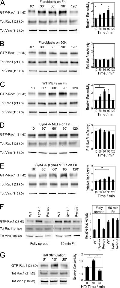
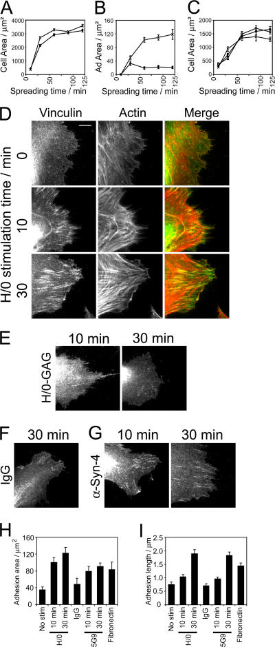
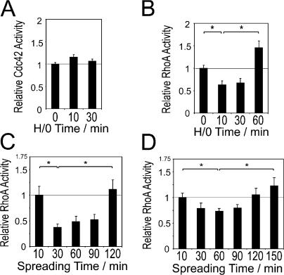
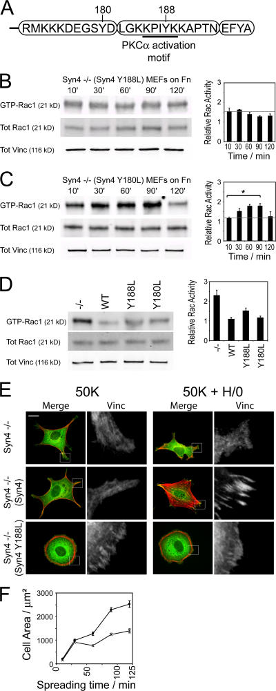
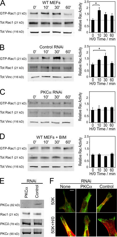
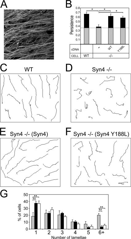
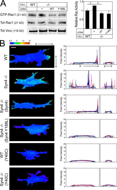
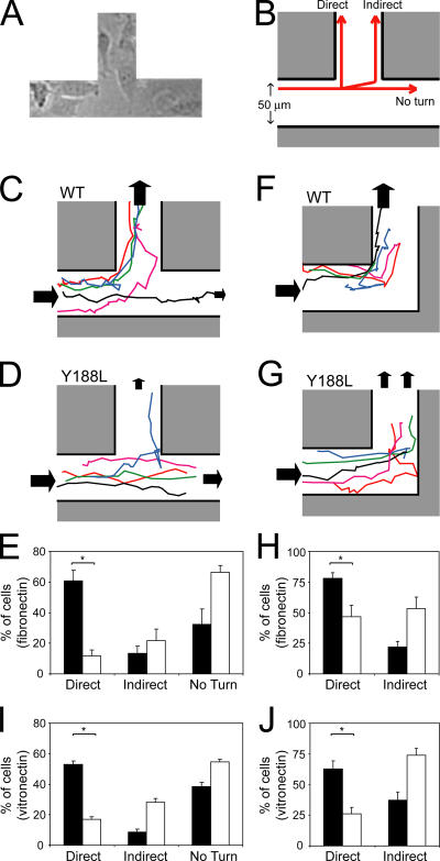
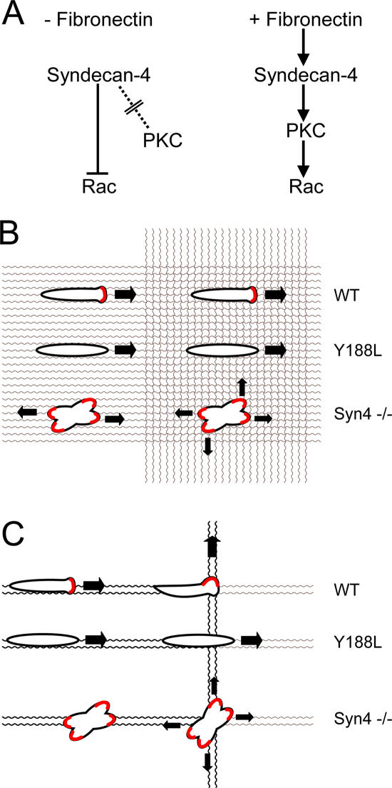
Cell migration in wound healing and disease is critically dependent on integration with the extracellular matrix, but the receptors that couple matrix topography to migratory behavior remain obscure. Using nano-engineered fibronectin surfaces and cell-derived matrices, we identify syndecan-4 as a key signaling receptor determining directional migration. In wild-type fibroblasts, syndecan-4 mediates the matrix-induced protein kinase Calpha (PKCalpha)-dependent activation of Rac1 and localizes Rac1 activity and membrane protrusion to the leading edge of the cell, resulting in persistent migration. In contrast, syndecan-4-null fibroblasts migrate randomly as a result of high delocalized Rac1 activity, whereas cells expressing a syndecan-4 cytodomain mutant deficient in PKCalpha regulation fail to localize active Rac1 to points of matrix engagement and consequently fail to recognize and respond to topographical changes in the matrix.


Figures

Figure 1.
Figure 1.
Engagement of syndecan-4 is essential for activation of Rac1 during adhesion to fibronectin. GTP-Rac1 levels during cell spreading or in response to H/0 were measured by effector pull-down assays in combination with quantitative Western blotting using fluorophore-conjugated antibodies. (A and B) Primary human fibroblasts were plated onto fibronectin (A) or 50K (B), and lysates were prepared after appropriate time periods. (C–E) The necessity of syndecan-4 expression for Rac1 regulation during spreading on fibronectin was tested using wild-type (C), syndecan-4–null (D), or syndecan-4–null transfected with full-length syndecan-4 cDNA MEFs (E). (F) Relative levels of GTP-Rac1 were directly compared between cell lines either fully spread (120 min) or during spreading on fibronectin for 60 min. (G) Rac1 activation in response to soluble H/0 in primary fibroblasts prespread on 50K. Equivalent loading between experiments was confirmed by blotting crude lysates for total Rac1 and vinculin. Axes are given in arbitrary units assigned according to the relative activity of fully spread cell lines. Each panel is representative of at least four separate experiments, and error bars indicate SEM. Asterisks indicate significant activation (*, P < 0.05).
Figure 2.
Figure 2.
Engagement of syndecan-4 drives the biphasic formation of adhesion complexes. The processes of spreading and adhesion complex formation were followed by staining fixed cells for vinculin and actin and measuring the cell area (A and C) or focal adhesion area (B and H) of 100 cells or the mean focal adhesion length (I) of 30 cells using ImageJ software. (A and B) Primary fibroblasts spreading on 50K (circles) or fibronectin (crosses). (C) Wild-type (crosses), syndecan-4–null (circles), or rescued (squares) MEFs spreading on fibronectin. (D–G) Adhesion complex formation in response to syndecan-4 engagement was followed in primary fibroblasts prespread on 50K for 2 h before stimulation with H/0 (D), a nonheparin-binding mutant of H/0 (E), nonimmune IgG (F), or 5G9 monoclonal antibody directed against the syndecan-4 extracellular domain (G). (H and I) Focal adhesion area (H) and mean focal contact length (I) of primary fibroblasts prespread on 50K for 2 h before stimulation with syndecan-4 ligands. Images and analyses are representative of experiments performed on six separate occasions. Error bars indicate SEM. Bar, 10 μm.
Figure 3.
Figure 3.
Engagement of syndecan-4 contributes toward but is not essential for the regulation of RhoA during adhesion to fibronectin. GTPase activity was measured by effector pull-down assays in combination with quantitative Western blotting using fluorophore-conjugated antibodies. (A and B) Primary fibroblasts were prespread on 50K for 2 h before measuring the activity of Cdc42 (A) or RhoA (B) in response to stimulation with H/0. (C and D) RhoA activity was measured during spreading on fibronectin (C) or 50K (D). Equivalent loading between experiments was confirmed by blotting crude lysates for total GTPase and vinculin. Each panel is representative of at least four separate experiments, and error bars indicate SEM. Asterisks indicate significant activation (P < 0.05).
Figure 4.
Figure 4.
The PKCα-binding motif of syndecan-4 mediates Rac1 regulation and adhesion complex formation. (A) Schematic representation of the syndecan-4 cytoplasmic domain. Tyr-188 is a key element of the PKCα-binding motif (Lim et al., 2003), and Tyr-180 was chosen as a negative control. (B and C) Syndecan-4–null MEFs expressing Y188L (B) or Y180L (C) mutant cDNAs were plated onto fibronectin, and GTP-Rac1 levels were measured by effector pull-down assays in combination with quantitative Western blotting using fluorophore-conjugated antibodies. (D) Relative levels of GTP-Rac1 in fully spread cells were compared between lines. Equivalent loading between experiments was confirmed by blotting crude lysates for total Rac1 and vinculin. Error bars indicate SEM, and asterisks indicate significant activation (P < 0.05). (E) Morphology of untransfected syndecan-4–null MEFs and MEFs expressing either wild-type or Y188L mutant cDNAs spread on 50K for 2 h before stimulation with H/0 for 60 min. Fixed cells were stained for vinculin (green) and actin (red). Boxes areas are magnified on the right. (F) The spreading profiles of MEFs expressing wild-type (crosses) or Y188L mutant (circles) syndecan-4 were followed by staining fixed cells for actin and measuring the cell area. All panels are representative of four separate experiments. Bar, 20 μm.
Figure 5.
Figure 5.
PKC activity is necessary for the regulation of Rac1 and adhesion complex formation. MEFs were prespread on 50K for 2 h before stimulation with H/0, and GTP-Rac1 levels were measured by effector pull-down assays in combination with quantitative Western blotting using fluorophore-conjugated antibodies. (A–D) Comparison of wild-type MEFS (A) with MEFs transfected with a control RNAi (B), an RNAi targeted against PKC (C), or treated with 200 nM BIM-1 for 30 min before and throughout stimulation (D). Equivalent loading between experiments was confirmed by blotting crude lysates for total Rac1 and vinculin. Error bars indicate SEM, and asterisks indicate significant activation (P < 0.05). (E) The effect of RNAi oligonucleotides on the expression of PKCα, Rac1, PKCδ, or PKCɛ. (F) Untransfected MEFs or MEFs transfected with PKCα-targeted or control RNAi were spread on 50K for 2 h and stimulated with H/0 for 60 min before fixing and staining for vinculin (green) and actin (red). All panels are representative of four separate experiments. Bar, 10 μm.
Figure 6.
Figure 6.
Expression of syndecan-4 determines the persistence of migration on cell-derived matrices. (A) Cell-derived matrices were generated by culturing human fibroblasts for 8 d before denuding the confluent fibroblasts and reseeding mutant MEF lines. (B) MEFs were seeded onto cell-derived matrices and were allowed to grow for 8 h before filming for 10 h. Persistence was determined by dividing the linear displacement of a cell by total distance migrated. Gray blocks represent the experimentally determined threshold for the random migration of cells on fibronectin-coated glass. (C–F) Migration tracks of MEFs over the 10-h filming period. The tracks of cells from three different fields of view have been compressed into each panel. (G) The number of lamellae present in syndecan-4–null MEFs (gray), reexpressing wild-type (black), or Y188L mutant (white) syndecan-4 were scored manually in all tracked cells at a single time point. Error bars indicate the SEM of 30 different cells, and asterisks indicate a significant difference in persistence (*, P < 0.05) or number of lamellae (**, P < 0.005). All panels are representative of four separate experiments.
Figure 7.
Figure 7.
Expression of syndecan-4 regulates the localized activity of Rac1. (A) MEF lines were allowed to spread on cell-derived matrices for 4 h before GTP-Rac1 levels were measured using effector pull-down assays in combination with quantitative Western blotting using fluorophore-conjugated antibodies. Error bars indicate SEM, and asterisks indicate significant activation (*, P < 0.01). Panels are representative of four separate experiments. (B) The distribution of Rac1 activity in situ was assessed by introducing a Raichu-Rac FRET probe and calculating the ratio of YFP/CFP emission upon excitation of CFP. Mean FRET intensity profiles were measured both parallel and perpendicular to the matrix fibrils. Panels are representative of 50 different cells, and the experiment was repeated on four separate occasions. Bar, 20 μm.
Figure 8.
Figure 8.
The PKCα-binding motif of syndecan-4 allows cells to turn in response to changes in the matrix environment. (A and B) MEFs were seeded onto fibronectin-coated gold stripes (A) and allowed to spread for 2 h before filming for 10 h and classifying turns as direct (retaining contact with the apex), indirect (forced by collision with the opposing wall), or no turn (B). (C–J) MEFs rescued with wild-type (C and F) or Y188L (D and G) syndecan-4 were tracked moving past a branch point with the option of turning (C and D) or toward a corner, where they were forced to turn (F and G). Cells migrating along gold stripes coated with fibronectin (E and H) or vitronectin (I and J) were scored for the ability to make direct, indirect, or no turn as they migrated past a junction (E and I) or toward a corner (H and J). 60 cells expressing wild-type (black bars) or Y188L mutant (white bars) syndecan-4 were tracked for each condition, and the experiment was repeated on three separate occasions. Error bars indicate the SEM, and asterisks indicate a significant difference (*, P < 0.001).
Figure 9.
Figure 9.
Engagement of syndecan-4 determines both the direction and persistence of migration. (A) Syndecan-4 limits Rac1 activity in the absence of matrix engagement and induces activation in response to fibronectin. (B and C) By constraining localized Rac1 activation to developing points of contact with the ECM (red), syndecan-4 coordinates migration along a fibronectin fibril. Consequently, wild-type fibroblasts migrate persistently over a meshwork of similar fibers (B) but follow the dominant strand when presented with a choice of paths (C). In contrast, syndecan-4–null cells protrude in multiple directions, rendering progression inefficient, whereas mutants in the PKCα-binding motif fail to respond to changes in matrix organization or follow the optimal path.

References

    1. Arthur, W.T., and K. Burridge. 2001. RhoA inactivation by p190RhoGAP regulates cell spreading and migration by promoting membrane protrusion and polarity. Mol. Biol. Cell. 12:2711–2720. - PMC - PubMed
    1. Bass, M.D., and M.J. Humphries. 2002. Cytoplasmic interactions of syndecan-4 orchestrate adhesion receptor and growth factor receptor signalling. Biochem. J. 368:1–15. - PMC - PubMed
    1. Bass, M.D., M.R. Morgan, and M.J. Humphries. 2007. Integrins and syndecan-4 make distinct, but critical, contributions to adhesion contact formation. Eur. Phys. J. E. Soft Matter. 3:372–376. - PMC - PubMed
    1. Bloom, L., K.C. Ingham, and R.O. Hynes. 1999. Fibronectin regulates assembly of actin filaments and focal contacts in cultured cells via the heparin-binding site in repeat III13. Mol. Biol. Cell. 10:1521–1536. - PMC - PubMed
    1. Burridge, K., and K. Wennerberg. 2004. Rho and Rac take center stage. Cell. 116:167–179. - PubMed

Publication types

MeSH terms