Comparative Study
. 2007 May 14;204(5):1227-35.
doi: 10.1084/jem.20070145. Epub 2007 May 7.

Lysosomal integral membrane protein 2 is a novel component of the cardiac intercalated disc and vital for load-induced cardiac myocyte hypertrophy

Affiliations
Comparative Study

Lysosomal integral membrane protein 2 is a novel component of the cardiac intercalated disc and vital for load-induced cardiac myocyte hypertrophy

Blanche Schroen et al. J Exp Med. .

Abstract

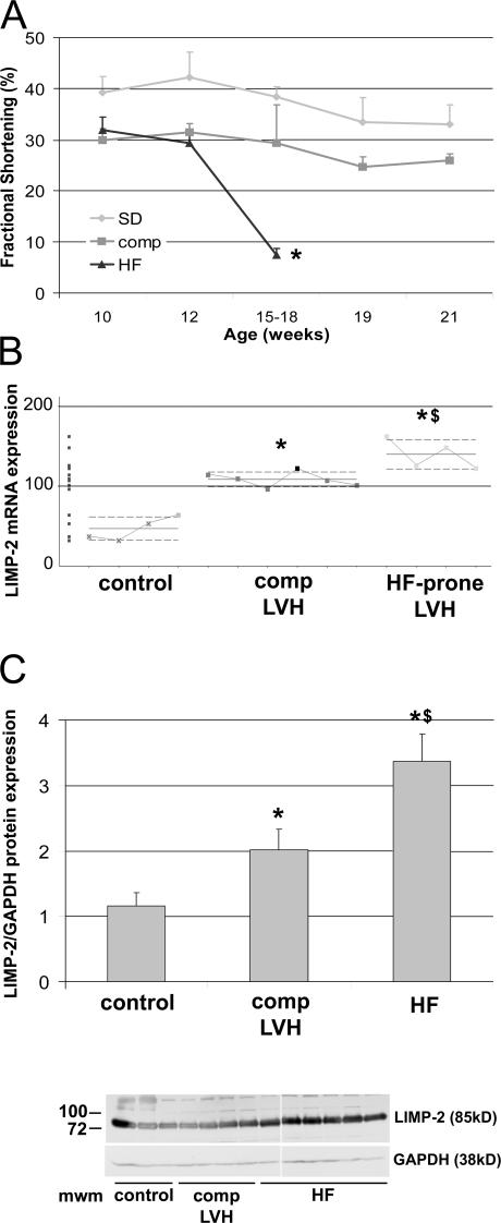
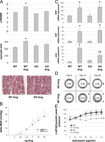
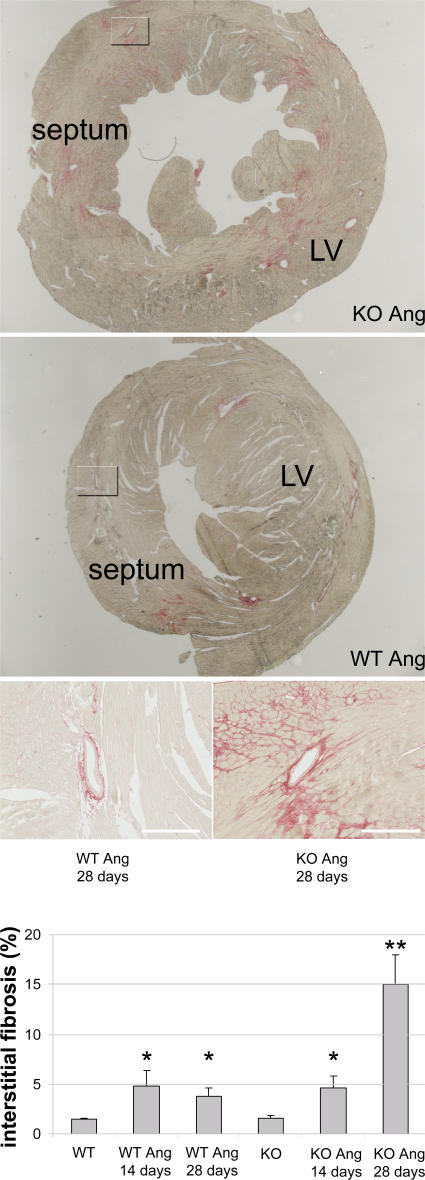
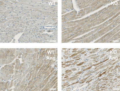
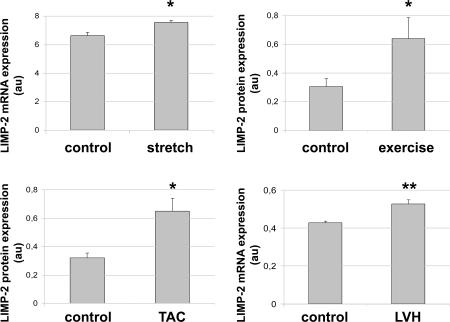
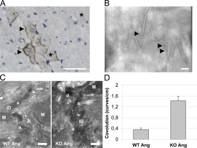
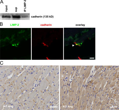
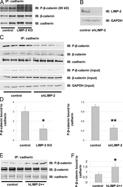
The intercalated disc (ID) of cardiac myocytes is emerging as a crucial structure in the heart. Loss of ID proteins like N-cadherin causes lethal cardiac abnormalities, and mutations in ID proteins cause human cardiomyopathy. A comprehensive screen for novel mechanisms in failing hearts demonstrated that expression of the lysosomal integral membrane protein 2 (LIMP-2) is increased in cardiac hypertrophy and heart failure in both rat and human myocardium. Complete loss of LIMP-2 in genetically engineered mice did not affect cardiac development; however, these LIMP-2 null mice failed to mount a hypertrophic response to increased blood pressure but developed cardiomyopathy. Disturbed cadherin localization in these hearts suggested that LIMP-2 has important functions outside lysosomes. Indeed, we also find LIMP-2 in the ID, where it associates with cadherin. RNAi-mediated knockdown of LIMP-2 decreases the binding of phosphorylated beta-catenin to cadherin, whereas overexpression of LIMP-2 has the opposite effect. Collectively, our data show that LIMP-2 is crucial to mount the adaptive hypertrophic response to cardiac loading. We demonstrate a novel role for LIMP-2 as an important mediator of the ID.


Figures

Figure 1.
Figure 1.
Increased expression of LIMP-2 in Ren-2 rats. An LV biopsy was taken at 10 wk of age, when Ren-2 rats exhibit comparable cardiac hypertrophy (reference 10) and fractional shortening cannot distinguish between rats that will progress to HF or those that will stay compensated (A). Between 15 and 18 wk of age, part of the Ren-2 rats progressed to HF, whereas the remainder stayed compensated until they were killed at 21 wk of age *, P < 10−5. In 10-wk-old hypertrophic Ren-2 rats, microarray analysis showed specific overexpression of LIMP-2 mRNA in failure-prone rats (HF-prone LVH; n = 4) as compared with the hypertrophied hearts that remained compensated (comp LVH; n = 6) and to controls (n = 4) (B). LIMP-2 protein was up-regulated in end-stage failing Ren-2 rats (HF; n = 9) as compared with compensated Ren-2 rats (comp; n = 6) and control rats (n = 6) (C). *, P < 0.05 versus control; **, P < 0.01 versus control; $, P < 0.05 versus comp; Mwm, molecular weight marker; au, arbitrary units.
Figure 2.
Figure 2.
AngII treatment in LIMP-2 KO (KO Ang) mice induces dilated cardiomyopathy. AngII increased LV weight and myocyte area in WT mice (n = 14) but not in KO mice (n = 14; *, P < 0.01 versus WT; n = 8) (A). Bars, 50 μm. LIMP-2 KO (n = 3) and WT (n = 4) mice showed comparable blood pressure responses to AngII (B). BNP, ANF, and aska mRNAs were induced in WT Ang and KO Ang (*, P < 0.05 vs. baseline; n = 4), but aska to a significantly lesser extend in KO Ang ($, P < 0.05 vs. WT Ang) (C). Echocardiography shows hypertrophied LV walls in WT Ang at days 14 and 28, whereas KOs did not show hypertrophy but were dilated (*, P < 0.005 vs. baseline and KO Ang; $, P < 0.005 vs. baseline and WT Ang; all groups, n ≥ 9) (D). β-adrenergic response to dobutamin was decreased in KO Ang mice (WT and KO, n = 4; WT Ang, n = 14; KO Ang, n = 9; *, P < 0.005) (E). LVW/BW, LV weight corrected for body weight.
Figure 3.
Figure 3.
After 4 wk of AngII treatment, LIMP-2 KO mice have massive interstitial fibrosis. Sirius red staining of LVs of 4-wk AngII-treated LIMP-2 KO (n = 4) and WT (n = 5) mice shows marked interstitial fibrosis in KO mice (*, P < 0.02 vs. WT Ang and KO baseline). The overview shows that this fibrosis was not uniformly distributed, but patchy. After 2 wk of AngII treatment, LIMP-2 KO and WT mice have similar induction of interstitial fibrosis, suggesting that the massive fibrosis after 4 wk is secondary. Both KO and WT mice treated with AngII show a similar degree of perivascular fibrosis. Bars, 250 μm.
Figure 4.
Figure 4.
AngII-treated LIMP-2 KO mice have a disturbed internal architecture. Desmin-stained cardiac myocytes of AngII-treated LIMP-2 KO mice have a disturbed internal structure, as shown by the higher and more capricious desmin expression in these mice. In AngII-treated KO mice, fibrotic areas were not taken into account because here myocyte suffering is evident. Bars, 250 μm.
Figure 5.
Figure 5.
LIMP-2 expression is up-regulated in other forms of mechanical loading. In neonatal rat cardiac myocytes, 6 h of stretch elevated LIMP-2 mRNA expression (n = 4 per group) (a). LIMP-2 expression was also up-regulated in hypertrophic myocardium from rats that had undergone exercise training for 10 wk (5 d per week; n = 6) as compared with nonhypertrophic control rats (n = 7) (b). After 1 h of transverse aortic banding in mice, LIMP-2 protein is elevated (c). LIMP-2 mRNA is up-regulated in hypertrophic patients with aortic stenosis (LVH; n = 20) as compared with nonhypertrophic control patients (n = 7) (d). *, P < 0.05 versus control; **, P < 0.01 versus control; LVH; LV hypertrophy.
Figure 6.
Figure 6.
LIMP-2 is present at the plasma membrane of cardiac myocytes and is important for ID function. Light (A) and immuno-electron (B) microscopy on a pressure-overloaded mouse, respectively. Rat LV tissue sections show LIMP-2 staining in intracellular compartments (*) and on plasma membranes of cardiac myocytes (arrows). Bars, 250 μm (A), respectively, 1 μm (B). Electron microscopy shows normal IDs in AngII-treated WT mice, whereas in AngII-treated LIMP-2 KO mice, the IDs have a higher degree of convolution, paralleled by the dilated cardiomyopathy in these mice (C). Bars, 2 μm. Degree of convolution was quantified (blinded) by counting the number of curves per centimeter ID on electron microscopic pictures and differed significantly (*, P < 10−5 vs. WT AngII; n = 15 for WT AngII and n = 15 for KO AngII) (D). M, mitochondrion; a, adherens junction; d, desmosome.
Figure 7.
Figure 7.
LIMP-2 regulates cadherin distribution. (A) LIMP-2 binds to cadherin in neonatal rat ventricular myocytes. LIMP-2 was immunoprecipitated (IP), and cadherin was immunoblotted (IB) in the total cell lysate (input), the supernatant (sup), and the precipitated protein lysate (IP). Part of the cadherin protein content of cardiac myocytes is bound by LIMP-2. (B) Representative tissue section of an HF patient was immunofluorescently stained with anti–pan-cadherin (red) and anti–LIMP-2 (green). The arrow shows colocalization of LIMP-2 and cadherin at the ID of cardiac myocytes. Bar, 50 μm. Tissue sections of AngII-treated LIMP-2 KO and WT LVs were immunostained with anti–pan-cadherin (C). In WT mice, the cadherin distribution is confined to the IDs yielding a regular appearance of cadherin, whereas in LIMP-2 KO mice, the localization of cadherin has lost the typical pattern produced by a strict location at the ID. Bars, 250 μm.
Figure 8.
Figure 8.
LIMP-2 regulates ID integrity by regulating the binding of phosphorylated β-catenin to cadherin. Immunoblot (IB) of lysates of neonatal rat cardiac myocytes that were treated either with shLIMP-2 or control shRNA (B). After 10 d of culture, cardiac myocytes show a 92% knockdown of LIMP-2 protein. Equal protein loading was confirmed by GAPDH. Representative immunoblots (IB) show diminished levels of P-β-catenin after IP with anti–pan-cadherin in LIMP-2 KO tissue lysates (A) as well as in shLIMP-2 cell lysates (C) as compared with control. P-β-catenin in total shLIMP-2 and control protein lysates was comparable (C). Quantification of immunoblots (n = 3 per group for control and LIMP-2 KO tissue lysates; n = 4 per group for control and shLIMP-2 cell lysates) is shown in D. Overexpression of LIMP-2 in neonatal rat cardiac myocytes increases levels of P-β-catenin bound to pan-cadherin (E and F). *, P < 0.05 versus control; **, P < 0.001 versus control.

References

    1. Opie, L.H. 2006. Controversies in cardiology. Lancet. 367:13–14. - PubMed
    1. Perriard, J.C., A. Hirschy, and E. Ehler. 2003. Dilated cardiomyopathy: a disease of the intercalated disc? Trends Cardiovasc. Med. 13:30–38. - PubMed
    1. Radice, G.L., H. Rayburn, H. Matsunami, K.A. Knudsen, M. Takeichi, and R.O. Hynes. 1997. Developmental defects in mouse embryos lacking N-cadherin. Dev. Biol. 181:64–78. - PubMed
    1. Ruiz, P., V. Brinkmann, B. Ledermann, M. Behrend, C. Grund, C. Thalhammer, F. Vogel, C. Birchmeier, U. Gunthert, W.W. Franke, and W. Birchmeier. 1996. Targeted mutation of plakoglobin in mice reveals essential functions of desmosomes in the embryonic heart. J. Cell Biol. 135:215–225. - PMC - PubMed
    1. Haegel, H., L. Larue, M. Ohsugi, L. Fedorov, K. Herrenknecht, and R. Kemler. 1995. Lack of beta-catenin affects mouse development at gastrulation. Development. 121:3529–3537. - PubMed

Publication types

MeSH terms