. 2007 Jun;88(Pt 6):1753-1760.
doi: 10.1099/vir.0.82666-0.

Type I feline coronavirus spike glycoprotein fails to recognize aminopeptidase N as a functional receptor on feline cell lines

Affiliations

Type I feline coronavirus spike glycoprotein fails to recognize aminopeptidase N as a functional receptor on feline cell lines

Charlotte Dye et al. J Gen Virol. 2007 Jun.

Abstract

There are two types of feline coronaviruses that can be distinguished by serology and sequence analysis. Type I viruses, which are prevalent in the field but are difficult to isolate and propagate in cell culture, and type II viruses, which are less prevalent but replicate well in cell culture. An important determinant of coronavirus infection, in vivo and in cell culture, is the interaction of the virus surface glycoprotein with a cellular receptor. It is generally accepted that feline aminopeptidase N can act as a receptor for the attachment and entry of type II strains, and it has been proposed that the same molecule acts as a receptor for type I viruses. However, the experimental data are inconclusive. The aim of the studies reported here was to provide evidence for or against the involvement of feline aminopeptidase N as a receptor for type I feline coronaviruses. Our approach was to produce retroviral pseudotypes that bear the type I or type II feline coronavirus surface glycoprotein and to screen a range of feline cell lines for the expression of a functional receptor for attachment and entry. Our results show that type I feline coronavirus surface glycoprotein fails to recognize feline aminopeptidase N as a functional receptor on three continuous feline cell lines. This suggests that feline aminopeptidase N is not a receptor for type I feline coronaviruses. Our results also indicate that it should be possible to use retroviral pseudotypes to identify and characterize the cellular receptor for type I feline coronaviruses.


Figures

Fig. 1.
Fig. 1.
Intracellular and cell surface expression of FCoV surface glycoproteins. (a) HEK 293T cells grown on coverslips were transfected with (i) pCAGGS/FCoVII-Str, (ii) pCAGGS/FCoVII-S, (iii) pCAGGS/FCoVI-Str, (iv) pCAGGS/FCoVI-S or (v) pCAGGS/Ta1 plasmid DNA. Cells were permeabilized and stained 48 h post-transfection using 23F4.4 mAb [(i), (ii) and (v)] or 210-70-FIP1 polyclonal serum [(iii) and (iv)] as the primary antibody and AlexaFluor-488 goat anti-mouse IgG [(i), (ii) and (v)] or FITC-conjugated goat anti-cat IgG [(iii) and (iv)] as the secondary antibody. Stained cells were viewed using a fluorescence microscope. (b) HEK 293T cells were transfected with (i) pCAGGS/FCoVII-Str, (ii) pCAGGS/FCoVII-S, (iii) pCAGGS/FCoVI-Str or (iv) pCAGGS/FCoVI-S, or plasmid DNA. Cells were trypsinized and stained 48 h post-transfection using the 23F4.4 mAb [(i) and (ii)] or the 210-70-FIP1 polyclonal serum [(iii) and (iv)] as the primary antibody and AlexaFluor-488 goat anti-mouse IgG [(i) and (ii)] or FITC-conjugated goat anti-cat IgG [(iii) and (iv)] as the secondary antibody. Cells were analysed for fluorescence by flow cytometry using the FACScan system.
Fig. 2.
Fig. 2.
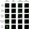
Cellular tropism of viral pseudotypes. CrFK (a, b, c and d), Fcwf-4 (e, f, g and h), FCKU (i, j, k and l), HEK 293T (m, n, o and p) and ST (q, r, s and t) cells were incubated with MLV(VSV-G) (a, e, i m and q), MLV(bald) (b, f, j, n and r), MLV(FCoVII-Str) (c, g, k, o and s) or MLV(FCoVI-Str) (d, h, l, p and t) viral pseudotypes. At 72 h post-incubation, the cells were analysed for GFP expression at 520 nm using a ×10 (a to h) or ×40 (i to t) objective on a Nikon Eclipse TS100 fluorescence microscope.
Fig. 3.
Fig. 3.
ELISA of FCoV type I S glycoprotein in MLV(FCoVI-Str) retroviral pseudotypes. ELISA microtitre plates were coated with cell culture supernatant containing a 1 : 32 dilution MLV(FCoVI-Str) retroviral pseudotypes diluted in coating buffer (▪), a 1 : 64 dilution MLV(FCoVI-Str) retroviral pseudotypes diluted in coating buffer (□), cell culture supernatant diluted in coating buffer (•) or coating buffer alone (○). Duplicate twofold dilutions of primary antibody (1 : 10–1 : 2560) were incubated with antigen-coated and uncoated wells and bound antibody was detected with AP-conjugated secondary antibody and pNPP. Plates were read at 405 nm.
Fig. 4.
Fig. 4.
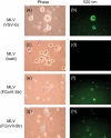
Transduction of feline adherent polymorphonuclear cells with MLV(FCoVI-Str) retroviral pseudotypes. Feline adherent polymorphonuclear cell cultures were incubated with MLV(VSV-G) (a and b), MLV(bald) (c and d), MLV(FCoVI-Str) (e and f) or MLV(FCoVII-Str) (g and h) viral pseudotypes. At 72 h post-incubation, the cells were visualized by microscopy (a, c, e and g) and analysed for GFP expression at 520 nm (b, d, f and h) using a Nikon Eclipse TS100 fluorescence microscope.

Similar articles

Cited by

References

    1. Addie, D. D. & Jarrett, O. (2001). Use of a reverse-transcriptase polymerase chain reaction for monitoring the shedding of feline coronavirus by healthy cats. Vet Rec 148, 649–653. - PubMed
    1. Addie, D. D., Schaap, I. A., Nicolson, L. & Jarrett, O. (2003). Persistence and transmission of natural type I feline coronavirus infection. J Gen Virol 84, 2735–2744. - PubMed
    1. Assavalapsakul, W., Smith, D. R. & Panyim, S. (2006). Identification and characterization of a Penaeus monodon lymphoid cell-expressed receptor for the yellow head virus. J Virol 80, 262–269. - PMC - PubMed
    1. Ausubel, F. M., Brent, R., Kingston, R. E., Moore, D. D., Seidman, J. G., Smith, J. A. & Struhl, K. (1987). Current Protocols in Molecular Biology. New York: Wiley.
    1. Benetka, V., Kubber-Heiss, A., Kolodziejek, J., Nowotny, N., Hofmann-Parisot, M. & Mostl, K. (2004). Prevalence of feline coronavirus types I and II in cats with histopathologically verified feline infectious peritonitis. Vet Microbiol 99, 31–42. - PMC - PubMed

Publication types

LinkOut - more resources