. 2007 May 15;104(20):8287-92.
doi: 10.1073/pnas.0611180104. Epub 2007 May 7.

Muller cells are living optical fibers in the vertebrate retina

Affiliations

Muller cells are living optical fibers in the vertebrate retina

Kristian Franze et al. Proc Natl Acad Sci U S A. .

Abstract

Although biological cells are mostly transparent, they are phase objects that differ in shape and refractive index. Any image that is projected through layers of randomly oriented cells will normally be distorted by refraction, reflection, and scattering. Counterintuitively, the retina of the vertebrate eye is inverted with respect to its optical function and light must pass through several tissue layers before reaching the light-detecting photoreceptor cells. Here we report on the specific optical properties of glial cells present in the retina, which might contribute to optimize this apparently unfavorable situation. We investigated intact retinal tissue and individual Müller cells, which are radial glial cells spanning the entire retinal thickness. Müller cells have an extended funnel shape, a higher refractive index than their surrounding tissue, and are oriented along the direction of light propagation. Transmission and reflection confocal microscopy of retinal tissue in vitro and in vivo showed that these cells provide a low-scattering passage for light from the retinal surface to the photoreceptor cells. Using a modified dual-beam laser trap we could also demonstrate that individual Müller cells act as optical fibers. Furthermore, their parallel array in the retina is reminiscent of fiberoptic plates used for low-distortion image transfer. Thus, Müller cells seem to mediate the image transfer through the vertebrate retina with minimal distortion and low loss. This finding elucidates a fundamental feature of the inverted retina as an optical system and ascribes a new function to glial cells.


Conflict of interest statement

The authors declare no conflict of interest.

Figures

Fig. 1.
Fig. 1.
Light transmission and reflection in the inner retina. (a) Experimental design to study light transmission through the inner retina. (Inset) Light emanating from a multimode optical fiber inserted into a freshly dissected eye simulates physiological illumination of the retina. The eye is opened at the posterior side, and all outer structures, including photoreceptor cells, are surgically removed. The laser light (λ = 543 nm) that is transmitted through the inner retina (NFL, nerve fiber layer; IPL, inner plexiform layer; INL, inner nuclear layer) is captured at the end of the prephotoreceptor light path with a confocal microscope. ONL, outer nuclear layer; ROS, photoreceptor outer segments. (b) Confocal transmission image of a living unstained retina. The brighter the signal, the more light is relayed to the corresponding area of the tissue. (c) Light reflection in the inner retina. Laser light is delivered via the microscope objective of an upright confocal microscope, and light scattered back from inner retinal layers is detected. (d) Confocal reflection image at the level of the IPL. The brighter the signal, the more light is reflected by the corresponding area. (Scale bar, 10 μm; also applies to b.)
Fig. 2.
Fig. 2.
Structures of low reflection are Müller cells. (a) Z-line reconstruction of reflection images of a living retina. The main scattering elements (bright) are the axon bundles and both plexiform layers. Low-reflecting tubular structures span the entire retina. (b) Living retinal slice preparation, visualizing Müller cells with the vital dye CellTracker orange (green) and synaptic elements in both plexiform layers (IPL and OPL) with the activity-dependent dye FM1–43 (red) (20). The levels of the inner and outer plexiform layers (IPL and OPL, respectively) and nerve fiber layer (NFL) are the same as in a. The asterisks indicate axon bundles in the NFL. (c and d) Overlay of light detected in reflection mode (purple) and the green fluorescence of the vital dye CellTracker green. (c) Z-line reconstruction of a confocal image stack. (d) Oblique optical section at the level of the red horizontal line in c. The dye-filled irregularly shaped Müller cell somata of the inner nuclear layer (INL) are visible in the left upper part. The central area shows Müller cell cross-sections in the IPL. In the lower right part, the Müller cell endfeet are visible, which enclose the ganglion cell somata in the ganglion cell layer (GCL). The lack of merging of the two colors, which would result in white areas, demonstrates that the dye filled exclusively those structures that showed low light reflection. (e–g) Confocal image at the IPL of a retinal whole mount fixed in 4% paraformaldehyde after exposure to the green vital dye and immunocytochemical labeling of vimentin (red), which in the retina is specific to Müller cells (17, 22). (e) Fluorescence of the vital dye. (f) Vimentin immunofluorescence. (g) Overlay of e and f. Colocalization of the red and green dyes results in yellow labeling. The observed complete colocalization means that the vital dye-filled and the immunoreactive cells are identical and thus identifies the low-reflecting tubular structures as Müller cells. [Scale bars: b, 10 μm (also applies to a); c–g, 25 μm.]
Fig. 3.
Fig. 3.
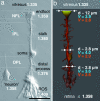
Müller cell shape, refractive properties, and light-guiding capability. (a) Nomarski differential interference contrast microscopy image of a dissociated guinea pig Müller cell with several adherent photoreceptor cells, including their outer segments (ROS) and a dissociated retinal neuron (bipolar cell) to the left. The refractive indices of the different cell sections are given. (b) Schematic illustration of a Müller cell in situ. The lighter the coloring of the Müller cell, the lower the refractive index. Typical diameters and the calculated V parameters for 700 nm (red) and 500 nm (blue) are indicated at the endfoot, the inner process, and the outer process. Although diameters and refractive indices change along the cell, its light-guiding capability remains fairly constant. (Scale bar, 25 μm.)
Fig. 4.
Fig. 4.
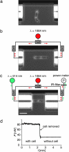
Demonstration of light guidance by individual Müller cells measured in a modified dual-beam laser trap. (a) A cell is floating freely between the ends of two optical fibers, which are aligned against a backstop visible at top. (b) The Müller cell is trapped, aligned, and stretched out by two counterpropagating near-infrared laser beams diverging from the optical fibers (42). (c) The fibers are brought in contact with the cell. Visible light (λ = 514 nm) emerges from the left (input) fiber and is collected and guided by the cell to the right (output) fiber. The fraction of visible light reentering the core of the output fiber is measured by a power meter, and the near-infrared light is blocked by an appropriate short-pass filter. (Scale bar, 50 μm.) (d) Typical time course of the power of visible light measured. When the cell is removed from the trap, it no longer prevents the light from diverging, and the measured power drops considerably. The ratio η = Pwith_cell/Pwithout_cell defines the relative guiding efficiency.

References

    1. Zernike F. Science. 1955;121:345–349. - PubMed
    1. Tuchin V. Tissue Optics. Bellingham, WA: SPIE Press; 2000.
    1. Maisel H. The Ocular Lens: Structure, Function, and Pathology. New York: Dekker; 1985.
    1. Snyder AW, Menzel R. Photoreceptor Optics. Berlin: Springer; 1975.
    1. Lee LP, Szema R. Science. 2005;310:1148–1150. - PubMed

Publication types