Comparative Study
. 2007 Oct 25;26(49):6979-88.
doi: 10.1038/sj.onc.1210508. Epub 2007 May 7.

The transcription factor ZEB1 (deltaEF1) promotes tumour cell dedifferentiation by repressing master regulators of epithelial polarity

Affiliations
Comparative Study

The transcription factor ZEB1 (deltaEF1) promotes tumour cell dedifferentiation by repressing master regulators of epithelial polarity

K Aigner et al. Oncogene. .

Abstract

Epithelial to mesenchymal transition (EMT) is implicated in the progression of primary tumours towards metastasis and is likely caused by a pathological activation of transcription factors regulating EMT in embryonic development. To analyse EMT-causing pathways in tumourigenesis, we identified transcriptional targets of the E-cadherin repressor ZEB1 in invasive human cancer cells. We show that ZEB1 repressed multiple key determinants of epithelial differentiation and cell-cell adhesion, including the cell polarity genes Crumbs3, HUGL2 and Pals1-associated tight junction protein. ZEB1 associated with their endogenous promoters in vivo, and strongly repressed promotor activities in reporter assays. ZEB1 downregulation in undifferentiated cancer cells by RNA interference was sufficient to upregulate expression of these cell polarity genes on the RNA and protein level, to re-establish epithelial features and to impair cell motility in vitro. In human colorectal cancer, ZEB1 expression was limited to the tumour-host interface and was accompanied by loss of intercellular adhesion and tumour cell invasion. In invasive ductal and lobular breast cancer, upregulation of ZEB1 was stringently coupled to cancer cell dedifferentiation. Our data show that ZEB1 represents a key player in pathologic EMTs associated with tumour progression.


Figures

Figure 1
Figure 1
RNAi-mediated knockdown of ZEB1 reactivates epithelial gene expression. (a) RNAi-mediated knockdown of ZEB1 and Snail1 in MDA-MB-231 cells. Transcript levels of ZEB1 and Snail1 were determined by real-time PCR using the TaqMan system with assay-on-demand (Applied Biosystems, Foster City, CA, USA). The relative expression of ZEB1 and Snail1 was normalized to β-actin using the standard curve method described by the manufacturer. Mean values of the relative transcript levels of three independent experiments are shown. Bars represent standard error. (b) E-cadherin protein expression levels after knockdown of ZEB1 (si-ZEB1) and Snail1 (si-Snail) in MDA-MB-231 and CAMA1 cells. si-Control, unspecific scrambled siRNA. Actin was included as loading control. (c) Knockdown of ZEB1 (si-ZEB1) causes transcriptional upregulation of epithelial polarity genes (see also Table 1). Actin was included as loading control. (d) E-cadherin, Crumbs3 (CRB3) and HUGL2 mRNA levels upon knockdown of ZEB1 (si-ZEB1) and Snail1 (si-Snail). Actin was included as loading control.
Figure 2
Figure 2
Cell polarity proteins are upregulated and partially redistributed to the plasma membrane upon ZEB1 depletion. (a) Immunolocalization of Crumbs3 (CRB3), HUGL2, PATJ and ZO1 in MDA-MB-231 cells treated with unspecific (si-Control) or ZEB1-specific (si-ZEB1) siRNA. (b) Crumbs3 (CRB3) and HUGL2 protein levels are upregulated upon knockdown of ZEB1 (si-ZEB1). si-Control, unspecific scrambled si-RNA. (c) Immunolocalization of Crumbs3 (CRB3) and HUGL2 in E-cadherin-expressing MDA-MB-231 cells (MDA-Ecad). (d) Transcript levels of Crumbs3 (CRB3), HUGL2 and PATJ in mock- and E-cadherin-transfected MDA-MB-231 cells. Transcript levels were determined by RT–PCR. Transcript levels in MCF7 cells were included as positive control, actin as loading control. Bars in (a) and (c), 10 μm.
Figure 3
Figure 3
ZEB1 directly represses the proximal promoter of cell polarity genes. (a) Scheme of the Crumbs3 promoter. E-boxes are indicated as vertical black bars. (b) ZEB1 and Snail repress the human Crumbs3 promoter. The relative repression of promoter fragments (see scheme in (a)) by ZEB1 and Snail1 compared to empty control vectors are shown. Mean values are derived from four independent experiments. Bars represent standard error. (c) ZEB1 and Snail1 repress the proximal HUGL2 promoter (−743/+135). Black vertical bars in the promoter scheme denote the relative positions of the E-boxes. (d) Upper panel: ZEB1 associates with the proximal Crumbs3 promoter at the chromatin level. ZEB1-specific (ZEB1) or unrelated goat Abs (Control) were used in ChIP experiments. Amplified human Crumbs3 promoter fragments are shown (ChIP1, −282/−29; ChIP2, −924/−646; ChIP3, −2284/−2037. Illustration in (a) shows the relative position of the amplified Crumbs3 promoter fragments. Amount of Crumbs3 promoter in input confirms equal loading of chromatin. Lower panel: knockdown of ZEB1 (si-ZEB1) reduces the interaction of ZEB1 with Crumbs3 promoter fragments. si-Control, unspecific scrambled si-RNA (e). Upper panel: ZEB1 associates with the PATJ promoter at the chromatin level. Illustration denotes the relative position of the two E-boxes (black vertical bars) as well as the amplified DNA fragment in ChIP (−335/−127). Lower panel: depletion of ZEB1 by siRNA (si-ZEB1) results in reduced precipitation of PATJ promoter fragments compared to control experiments (scrambled si-Control).
Figure 4
Figure 4
ZEB1 and Snail depletion reduced the motility of MDA-MB-231 cells in vitro. MDA-MB-231, treated with control or ZEB1 and Snail-specific siRNAs for 3 days, and MDA-MB-231 cells stably transfected with empty or E-cadherin-expressing plasmids, were seeded on Transwell filter inserts and migrating cells quantified after 24h by Hoechst staining and fluorescence microscopy.
Figure 5
Figure 5
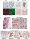
ZEB1 expression in colon and breast tumours correlates with cancer cell dedifferentiation. (a) Serial tumour sections were immunohistochemically stained for ZEB1, E-cadherin or cytokeratin (brown). In addition, single sections were double stained for ZEB1 and E-cadherin and subjected to immunofluorescence analysis (ZEB1 in red, E-cadherin in green). ZEB1 and HUGL2 represent immunohistochemical double stainings (ZEB1, brown; HUGL2, violet; two different magnifications of adjacent regions are shown). Note that differentiated areas (arrows) stain strongly for E-cadherin, cytokeratin and HUGL2 but do not express significant levels of ZEB1. Tumour-associated stroma cells express high amounts of nuclear ZEB1 (arrowheads). (b) ZEB1 induces EMT-like cell conversions at the invasive front of colorectal adenocarcinomas. Sections were double stained for ZEB1 (brown) and cytokeratin (violet). At the tumour–host interface ZEB1-positive tumour cells invade the host stroma but retain residual cytokeratin expression (see arrows in left panel: ZEB1, brown; cytokeratin, violet). Small cancer cell cluster or single isolated tumour cells express ZEB1 and often contain perinuclear and/or cytoplasmic cytokeratin remnants (see arrows in right panel: ZEB1, brown; cytokeratin, violet). (c) Expression of ZEB1 in breast tumours correlates with impaired cancer cell differentiation. Invasive ductal and lobular breast cancer specimens were double stained for ZEB1 (brown) and cytokeratin (violet). In differentiated areas of ductal tumours, ZEB1 is expressed in many tumour-associated stroma cells (Ductal-Diff., arrowhead), whereas the tumour lacks ZEB1-specific staining (Ductal-Diff., arrow). Dedifferentiated ductal compartments strongly express ZEB1 (Ductal-Dediff. and Mixed, arrows). In lobular cancer ZEB1 is highly expressed in large areas of the tumour (Lobular). Bars 20 and 80 μm.

References

    1. Batlle E, Sancho E, Franci C, Dominguez D, Monfar M, Baulida J, et al. The transcription factor snail is a repressor of E-cadherin gene expression in epithelial tumour cells. Nat Cell Biol. 2000;2:84–89. - PubMed
    1. Bilder D. Epithelial polarity and proliferation control: links from the Drosophila neoplastic tumor suppressors. Genes Dev. 2004;18:1909–1925. - PubMed
    1. Brabletz T, Jung A, Reu S, Porzner M, Hlubek F, Kunz-Schughart LA, et al. Variable beta-catenin expression in colorectal cancers indicates tumor progression driven by the tumor environment. Proc Natl Acad Sci USA. 2001;98:10356–10361. - PMC - PubMed
    1. Brabletz T, Jung A, Spaderna S, Hlubek F, Kirchner T. Opinion: migrating cancer stem cells – an integrated concept of malignant tumour progression. Nat Rev Cancer. 2005;5:744–749. - PubMed
    1. Brumby AM, Richardson HE. Using Drosophila melanogaster to map human cancer pathways. Nat Rev Cancer. 2005;5:626–639. - PubMed

Publication types

MeSH terms