. 2007 Jun;4(6):511-6.
doi: 10.1038/nmeth1048. Epub 2007 May 7.

A semisynthetic epitope for kinase substrates

Affiliations

A semisynthetic epitope for kinase substrates

Jasmina J Allen et al. Nat Methods. 2007 Jun.

Abstract

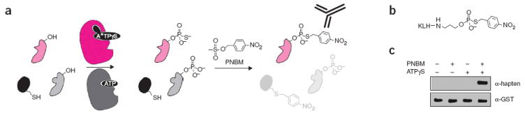
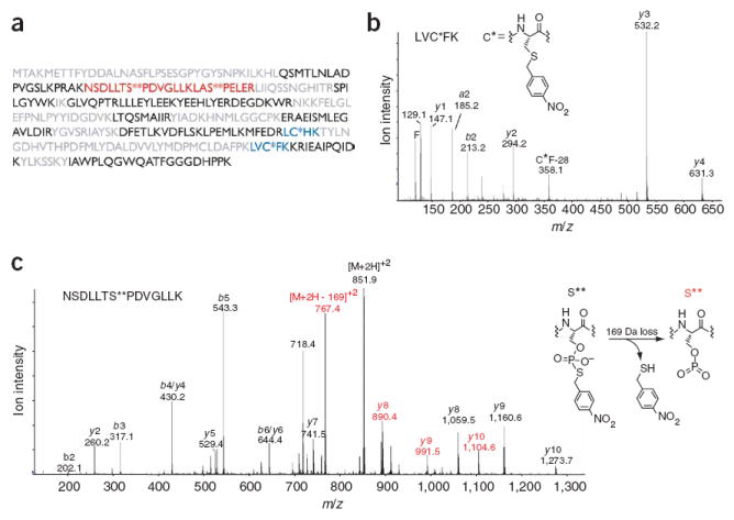
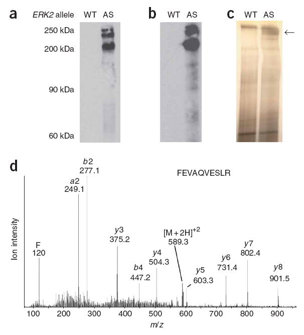
The ubiquitous nature of protein phosphorylation makes it challenging to map kinase-substrate relationships, which is a necessary step toward defining signaling network architecture. To trace the activity of individual kinases, we developed a semisynthetic reaction scheme, which results in the affinity tagging of substrates of the kinase in question. First, a kinase, engineered to use a bio-orthogonal ATPgammaS analog, catalyzes thiophosphorylation of its direct substrates. Second, alkylation of thiophosphorylated serine, threonine or tyrosine residues creates an epitope for thiophosphate ester-specific antibodies. We demonstrated the generality of semisynthetic epitope construction with 13 diverse kinases: JNK1, p38alpha MAPK, Erk1, Erk2, Akt1, PKCdelta, PKCepsilon, Cdk1/cyclinB, CK1, Cdc5, GSK3beta, Src and Abl. Application of this approach, in cells isolated from a mouse that expressed endogenous levels of an analog-specific (AS) kinase (Erk2), allowed purification of a direct Erk2 substrate.


Conflict of interest statement

COMPETING INTERESTS STATEMENT The authors declare competing financial interests: details accompany the full-text HTML version of the paper at www.nature.com/naturemethods.

Figures

Figure 1
Figure 1
Strategy for labeling individual kinase substrates. (a) Reaction sequence for affinity tagging AS kinase substrates. First, an AS kinase (magenta) uses N6-alkylated ATPγS (A*TPγS) to thiophosphorylate its substrates (pale magenta). In a second step, alkylation with PNBM yields thiophosphate esters and thioethers. Only AS kinase substrates are recognized by α-hapten–IgG. (b) Structure of the hapten conjugate used to elicit thiophosphate ester specific antibodies. (c) JNK1 was incubated with c-Jun–GST and combinations of ATPγS and PNBM as indicated; reaction products were analyzed by western blot with α-hapten–IgG. α-GST–IgG confirms equal loading. Full-length blots are available in Supplementary Figure 1b.
Figure 2
Figure 2
Mass spectrometric analysis of thiophosphorylated and alkylated c-Jun–GST. (a) Sequence coverage of c-Jun-GST. Residues in black were identified by automated database searching of the unmodified peptides, blue indicates peptides that contain a modified cysteine, red indicates thiophosphate ester–containing peptides, and residues in gray were not observed. (b) Tandem mass spectrum of a thioether–containing peptide. C* indicates a nitrobenzyl-modified cysteine. (c) Tandem mass spectrum of a peptide containing a modified serine (S**), which corresponds to a site of JNK1 phosphorylation. A 169-Da loss, consistent with the depicted elimination, was observed from the doubly charged molecular ion and also gave rise to an additional y-ion series. Ions that have undergone this loss are labeled in red.
Figure 3
Figure 3
α-hapten-IgG detection of thiophosphate-esterified kinase substrates. Each blot is labeled with the corresponding kinase and its preferred phosphorylation consensus motif. A description of the conditions for each kinase reaction is available in the Supplementary Methods; full-length blots are available in Supplementary Figure 1c.
Figure 4
Figure 4
A*TPγS analog orthogonality and acceptance by AS kinases. (a) Chemical structure of A*TPγS, R indicates the site of N6 modification. (b) Kinase reactions with ATPγS and A*TPγS analogs. Following PNBM alkylation, the labeled substrates were detected with α-hapten-IgG western blot analysis, full-length blots are available in Supplementary Figure 1d. WT, wild type.
Figure 5
Figure 5
Labeling, immunoprecipitation and identification of AS kinase substrates. (a) MEFs expressing either wild-type (WT) or AS Erk2 were permeabilized, incubated with A*TPγS, alkylated and analyzed by western blot (RmAb α-hapten–IgG). (b) Western blot analysis of the same samples as in a after immunoprecipitation. (c) Silver stain gel of immunoprecipitates in b. (d) Liquid chromatography–tandem mass spectrometry spectrum of a tryptic peptide, FEVAQVESLR, from the indicated band in c. Full-length blots and silver-stained gels are available in Supplementary Figure 1e.

References

    1. Berwick DC, Tavare JM. Identifying protein kinase substrates: hunting for the organ-grinder’s monkeys. Trends Biochem Sci. 2004;29:227–232. - PubMed
    1. Ptacek J, Snyder M. Charging it up: global analysis of protein phosphorylation. Trends Genet. 2006;22:545–554. - PubMed
    1. Ptacek J, et al. Global analysis of protein phosphorylation in yeast. Nature. 2005;438:679–684. - PubMed
    1. Morrison DK, Davis RJ. Regulation of MAP kinase signaling modules by scaffold proteins in mammals. Annu Rev Cell Dev Biol. 2003;19:91–118. - PubMed
    1. Biondi RM, Nebreda AR. Signalling specificity of Ser/Thr protein kinases through docking-site-mediated interactions. Biochem J. 2003;372:1–13. - PMC - PubMed

Publication types

MeSH terms