. 2007 Jun;4(6):501-9.
doi: 10.1038/nmeth1041. Epub 2007 May 7.

Generation of functional hemangioblasts from human embryonic stem cells

Affiliations

Generation of functional hemangioblasts from human embryonic stem cells

Shi-Jiang Lu et al. Nat Methods. 2007 Jun.

Abstract

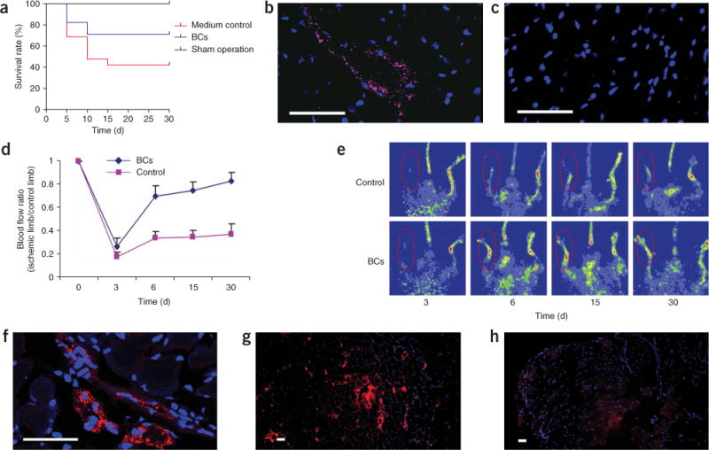
Recent evidence suggests the existence of progenitor cells in adult tissues that are capable of differentiating into vascular structures as well as into all hematopoietic cell lineages. Here we describe an efficient and reproducible method for generating large numbers of these bipotential progenitors-known as hemangioblasts-from human embryonic stem (hES) cells using an in vitro differentiation system. Blast cells expressed gene signatures characteristic of hemangioblasts, and could be expanded, cryopreserved and differentiated into multiple hematopoietic lineages as well as into endothelial cells. When we injected these cells into rats with diabetes or into mice with ischemia-reperfusion injury of the retina, they localized to the site of injury in the damaged vasculature and appeared to participate in repair. Injection of the cells also reduced the mortality rate after myocardial infarction and restored blood flow in hind limb ischemia in mouse models. Our data suggest that hES-derived blast cells (hES-BCs) could be important in vascular repair.


Conflict of interest statement

COMPETING INTERESTS STATEMENT

The authors declare competing financial interests: details accompany the full-text HTML version of the paper at www.nature.com/naturemethods.

Figures

Figure 1
Figure 1
Characterization of hES-BCs. (a) Phase-contrast images of a blast colony (left), secondary EBs (middle) and hES-BCs with Wright-Giemsa staining (right). (b) Phase-contrast images of Wright-Giemsa–stained cells from CFU-erythrocytes (left), cells from CFU-granulocytes (cells that form hematopoietic colonies with granulocytes, middle) and cells from CFU-macrophages (cells that form hematopoietic colonies with macrophages, right). (c) Immunostaining of cells from CFU-erythrocytes with CD235a antibody (left), cells from CFU-granulocytes with CD13 antibody (middle) and cells from CFU-GEMMs with CD45 antibody (right). (d) Analysis of endothelial cells derived from purified hES-BCs: Ac-LDL uptake (red) and immunostaining of vWF (green; left), uptake of Ac-LDL (red) and immunostaining of VE-cadherin (green; middle); immunostaining of vWF (green) and CD31 (red; right). DAPI (blue) staining of cell nuclei. Scale bars, 50 μm.
Figure 2
Figure 2
Clonogenicity of blast colonies. (a,b) Phase-contrast (a) and fluorescence (b) images of two blast colonies developed in a mixture of WA01-GFP and MA01 EBs, which demonstrated the clonal origin of blast colonies. (c) Expansion of a single blast colony in liquid culture; both hematopoietic and endothelial lineages were observed. Scale bars, 100 μm.
Figure 3
Figure 3
Repair of ischemic retinal vasculature in a mouse after injection of hES-BCs. (a–e) Mice undergoing ischemia-reperfusion injury were injected cells either intravitreally (a,b) or intravenously (c–e) with fluorescently labeled hES-BCs. Merged images from the ischemic eye of the same mouse 1 d after intravitreal hES-BC cell administration of fluorescently labeled hES-BCs (green channel) (a), or a typical control (uninjured) eye 1 d after saline injection (b). Separate green (hES-BCs) and red channels are shown in the insets. Merged images of the ischemic eyes 2 d (c) and 7 d (d) after systemic hES-BC cell administration and of the uninjured eye of the same mouse (e). (f) hES-BC–derived endothelial cells colocalize to existing injured vasculature in cross-sections of mouse eyes that underwent ischemia-reperfusion injury. High-magnification view of a vascular lumen in the ganglion cell layer adjacent to the inner limiting membrane (lower left) shows lumen surrounded by resident endothelial cells (CD31; green) and hES-BC–derived endothelial cells (human nuclear antigen, red) and the colocalized fluorescence staining of CD31 and human nuclear antigen (yellow). Upper right and left panels are the separate red (human nuclear antigen) and green (CD31) channels used to make composite image. Lower right panel, low magnification of the same region, with the box showing area depicted in all panels. V, vitreous; IPL, inner plexiform layer; RPE, retinal pigment epithelial cell layer; Ch, choroid. Scale bars, 100 μm in a–e (225 μm in insets), and 25 μm in f.
Figure 4
Figure 4
Incorporation of hES-BCs into the retinal vasculature of diabetic rats. (a,b) Merged images from the retinal vasculature of two separate diabetic rats 2 d after intravitreal hES-BC administration, showing extensive hES-BC cell incorporation into both large and small vessels. (c) Merged image (green and red channels) from a control (nondiabetic) rat 2 d after hES-BC administration, showing that hES-BCs did not incorporate into vasculature and formed a sheet that lay atop the retina. (d,e) Eye sections from diabetic rats 2 d after intravitreal hES-BC injection, stained with CD31 (green) and human nuclear antigen (red) antibodies, showing colocalized staining of CD31 (green) and human nuclear antigen (red) in endothelial cells lining vessel lumens in the ganglion cell layer of the retina. Arrows, colocalized staining (yellow) of CD31 (green) and human nuclear antigen (red). Insets, separate green and red channels. (f) A section from a nondiabetic control rat 2 d after intravitreal blast cell injection, which is negative for human nuclear antigen staining, but is positive for endothelium staining (CD31, green; arrows). Arrows, positive staining of CD31 (green), but negative for human nuclear antigen (red). Scale bars, 100 μm (a–c), and 25 μm (d–f).
Figure 5
Figure 5
Endothelial differentiation in infarcted heart and in ischemic hind limb muscle after injection of hES-BCs. (a–c) Differentiation of hES-BCs in infarcted hearts: survival curves of mice treated with sham operation, medium control and hES-BCs (a); confocal image of infarcted myocardium section 4 weeks after injection of hES-BCs, immunostained with human-specific vWF antibody (pink) and DAPI (nuclei, blue) (b); confocal image of infarcted myocardium section from control mouse immunostained with human-specific vWF antibody (pink) and DAPI (nuclei, blue), showing no stain of human vWF (c). (d–h) Differentiation of hES-BCs in ischemic hind limb muscles. Restoration of blood flow in surgically induced ischemic limbs by hES-BCs (d). Hind limb blood flow monitored serially for 3–30 d after ligation in mouse receiving 6 × 10⁵ hES-BCs and in mouse receiving medium only. Blood flow is calculated as the ratio of flow in the ischemic limb to that in the non-ischemic limb of the same animal. Laser doppler blood flow images of controls (medium) and ischemic animals injected with blast cells (n = 6 for each group) (e). Confocal microscopy image (f) and regular microscopy image (g) of ischemic hind limb muscle sections 4 weeks after injection of hES-BCs, immunostained with human-specific vWF antibody (red) and DAPI (nuclei, blue); an ischemic hind limb muscle section from a control mouse 4 weeks after PBS injection, immunostained with human specific vWF antibody (red) and DAPI (nuclei, blue), showing no stain of human vWF (h). Scale bars, 40 μm.

References

    1. Rafii S, Lyden D. Therapeutic stem and progenitor cell transplantation for organ vascularization and regeneration. Nat Med. 2003;9:702–712. - PubMed
    1. Grant MB, et al. Adult hematopoietic stem cells provide functional hemangioblast activity during retinal neovascularization. Nat Med. 2002;8:607–612. - PubMed
    1. Bailey AS, et al. Transplanted adult hematopoietic stems cells differentiate into functional endothelial cells. Blood. 2004;103:13–19. - PubMed
    1. Cogle CR, et al. Adult human hematopoietic cells provide functional hemangioblast activity. Blood. 2004;103:133–135. - PubMed
    1. Otani A, et al. Bone marrow-derived stem cells target retinal astrocytes and can promote or inhibit retinal angiogenesis. Nat Med. 2002;8:1004–1010. - PubMed

Publication types