. 2007 May 21;96(10):1595-604.
doi: 10.1038/sj.bjc.6603755. Epub 2007 May 8.

Non-genomic action of resveratrol on androgen and oestrogen receptors in prostate cancer: modulation of the phosphoinositide 3-kinase pathway

Affiliations

Non-genomic action of resveratrol on androgen and oestrogen receptors in prostate cancer: modulation of the phosphoinositide 3-kinase pathway

D A Benitez et al. Br J Cancer. .

Abstract

Prostate cancer represents a major concern in human oncology and the phytoalexin resveratrol (RES) inhibits growth and proliferation of prostate cancer cells through the induction of apoptosis. In addition, previous data indicate that in oestrogen-responsive human breast cancer cells, RES induces apoptosis by inhibition of the phosphoinositide-3-kinase (PI3K) pathway. Here, using androgen receptor (AR)-positive LNCaP and oestrogen receptor alpha (ERalpha)-expressing PC-3 prostate tumour cells, we have analysed whether the antiproliferative activity of RES takes place by inhibition of the AR- or ERalpha-dependent PI3K pathway. Although RES treatment (up to 150 microM) decreased AR and ERalpha protein levels, it did not affect AR and ERalpha interaction with p85-PI3K. Immunoprecipitation and kinase assays showed that RES inhibited AR- and ERalpha-dependent PI3K activities in LNCaP and PC-3, respectively. Consistently, lower PI3K activities correlated with decreased phosphorylation of downstream targets protein kinase B/AKT (PKB/AKT) and glycogen synthase kinase-3 (GSK-3). GSK-3 dephosphorylation could be responsible for the decreased cyclin D1 levels observed in both cell lines. Importantly, RES markedly decreased PKB/AKT phosphorylation in primary cultures from human prostate tumours, suggesting that the mechanism proposed here could take place in vivo. Thus, RES could have antitumoral activity in androgen-sensitive and androgen-non-sensitive human prostate tumours by inhibiting survival pathways such as that mediated by PI3K.


Figures

Figure 1
Figure 1
RES does not significantly affect p85/PI3K but decreases AR and ERα levels in LNCaP and PC-3 cells. LNCaP (A, C) and PC-3 (B, D) were left untreated (0, DMSO) or treated with 1, 10, 50, 100 or 150 μM RES for 36 h. Total protein extracts were obtained and analysed for p85/PI3K (A, B), AR (C) or ERα (D) protein expression by Western immunobloting using specific antibodies. The level of β-actin was also determined to account for protein quantitation and equal loading. p85/PI3K, AR and ERα expression were quantitated and normalised by β-actin in at least three different cultures. Data shown are mean±s.e. The difference with respect to untreated cultures (DMSO) is significant at P<0.05 (*).
Figure 2
Figure 2
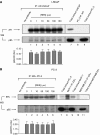
Steroid receptors AR and ERα interact with p85/PI3K in LNCaP and PC-3 prostate cancer cells and such interaction is not altered by RES treatment. LNCaP (A) and PC-3 (B), growing in complete medium, were treated with the indicated concentrations of RES for 36 h and 1 mg total cell extracts immunoprecipitated (IP) with anti-AR- or anti-ERα-specific antibodies, respectively. The amount of p85/PI3K associated to each receptor was determined in the immunoprecipitates by Western immunobloting (WB) (lower blots in A and B, lanes 1–6). For quantitation, immunoprecipitated p85 was normalised by the amount of AR (A) or ERα (B) at each concentration of RES in three different cultures (lanes 1–6). For LNCaP cells (A) total cell extract was used as a positive control for AR and p85 expression (lane 7), whereas negative controls for this cell line included AR immunoprecipitation (lane 8) and total extract (lane 9) from androgen-insensitive PC-3 cells. For PC-3 (B), positive controls for ERα and p85 expression included total cell extracts from this cell line (lane 7) and from human breast tumour MCF-7 cells (lane 9); for ERα association to p85, immunoprecipitations were carried out in oestrogen-responsive MCF-7 (lower blot in B, lane 8). Negative controls were also included for ERα expression (lane 11) and for its interaction with p85 (lane 10) using oestrogen-unresponsive human breast tumour MDA-MB-231 cells. As an additional negative control, immunoprecipitations were carried out in the absence of AR or ERα antibodies (no antibody in A and B).
Figure 3
Figure 3
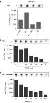
The AR-associated PI3K activity is modulated by RES in androgen-sensitive LNCaP cells. (A) LNCaP cells were cultured for 5 days in steroid-depleted medium and then treated with DMSO (control), 10−9M of the AR agonist DHT (30 min), 10−9M DHT (30 min) plus 10−4M of the AR antagonist Bic (30 min) or 150 μM RES (30 min). Aliquots of 1 mg total cell extract were immunoprecipitated with an AR-specific antibody and PI3K activity determined in the immunoprecipitates using PI as substrate. (B) PI3K activity was determined in AR immunoprecipitates obtained from cultures grown in steroid-depleted medium and left untreated (DMSO) or treated with 1, 10, 50, 100 or 150 μM RES for 36 h. Some cultures were treated with 20 μM of the PI3K inhibitor LY294002 (LY). (C) PI3K activity was also determined in AR immunoprecipitates from cultures grown in complete medium and treated with 0 (DMSO), 1, 10, 50, 100 or 150 μM RES. Androgen-insensitive PC-3 cells (PC3) were used as negative control. A representative experiment out of two is shown.
Figure 4
Figure 4
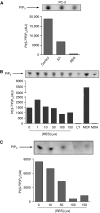
RES modulates the ERα-associated PI3K activity in androgen-insensitive PC-3 cells. (A) PC-3 cells were cultured in steroid-depleted medium for 5 days and then treated with DMSO (control), 1 μM of the ERα antagonist ICI 182,780 (30 min) or 150 μM RES (30 min). Total cell extracts were prepared and 1 mg protein immunoprecipitated with an ERα-specific antibody. ERα-associated PI3K activity was determined in the immunoprecipitates using PI as substrate. (B) PC-3 cells growing in steroid depleted medium for 5 days were left untreated (DMSO) or treated with 1, 10, 50, 100 or 150 μM RES for 36 h and PI3K activity determined in ERα immunoprecipitates. Cells were also treated with 20 μM of the PI3K inhibitor LY294002 (LY). As positive and negative controls, experiments were performed in ERα immunoprecipitates from ERα-positive MCF-7 and ERα-negative MDA-MB-231 breast cancer cells, respectively. (C) PC-3 cells were grown in complete medium and left untreated (DMSO) or treated with 10, 50, 100 or 150 μM RES for 36 h. PI3K activity was determined in ERα immunoprecipitates using PI as substrate. A representative experiment from two is shown.
Figure 5
Figure 5
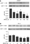
RES inhibition of the PI3K activity in LNCaP and PC-3 cells resulted in decreased PKB/AKT phosphorylation. LNCaP (A) and PC-3 (B) cells were left untreated (DMSO) or treated with 1, 10, 50, 100 or 150 μM RES for 36 h in complete medium. Protein extracts were analysed by Western immunobloting to detect the phosphorylated (pAKT) and the total PKB/AKT protein (tAKT). Data were quantitated by normalising pPKB/AKT by tPKB/AKT (pAKT/tAKT) and the results plotted against the concentration of RES. Data shown are mean±s.e. from three different cultures. The differences with respect to control cultures (DMSO) are significant at P<0.05 (*) or P<0.01 (**).
Figure 6
Figure 6
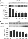
RES induces a pattern of GSK-3 phosphorylation in LNCaP and PC-3 cells that closely resembles that of PKB/AKT. LNCaP (A) and PC-3 (B) were treated with DMSO, 1, 10, 50, 100 or 150 μM RES for 36 h in complete medium. Total cell extracts were analysed for phosphorylated (pGSK3) and total GSK-3 (tGSK3) by Western immunobloting using specific antibodies. Phospho-GSK-3 was normalised by total GSK-3 protein at each concentration of RES. Data shown are mean±s.e. from at least three different experiments. The differences are statistically significant with respect to control values (DMSO) at P<0.05 (*) or P<0.01 (**).
Figure 7
Figure 7
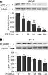
The GSK-3 target protein cyclin D1 is affected by RES in LNCaP and PC-3 cells with a pattern similar to that of PKB/AKT. LNCaP (A) and PC-3 (B) cells were left untreated (DMSO) or treated with 1, 10, 50, 100 or 150 μM RES for 36 h in complete medium. Protein extracts were analysed for cyclin D1 levels by Western immunobloting using a specific antibody. The expression of β-actin was also determined to account for equal loading and protein quantitation. Cyclin D1 was normalised by β-actin and the results plotted for each concentration of RES. Data are shown as mean±s.e. from three different cultures. The differences with respect to control cultures (DMSO) are significant at P<0.05 (*) or P<0.01 (**).
Figure 8
Figure 8
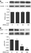
The effects of RES on p85/PI3K and PKB/AKT phosphorylation in cultured human primary prostate tumour cells closely resemble those observed in LNCaP and PC-3 cells. A total of seven biopsies from patients scheduled for radical prostatectomy were used to obtain primary cultures of prostate epithelial tumour cells. Cells were characterised as having a transformed phenotype as previously were indicated (Sanchez et al, 2005; Castellon et al, 2006, 2005). Cultures treated with DMSO, 10, 50 or 100 μM RES for 36 h in complete medium. (A) Total protein extracts were analysed for p85/PI3K expression by Western immunobloting and the results normalised by β-actin levels. (B) Phosphorylated (pAKT) and total PKB/AKT (tAKT) were also determined by Western immunobloting and the amount of active protein (pAKT) normalised by the total PKB/AKT (tAKT). Data are shown as mean±s.e. The differences with respect to untreated control cultures are significant at P<0.01 (**).
Figure 9
Figure 9
Proposed mechanism for RES induced apoptosis in LNCaP and PC-3 human prostate tumour cells. Concentrations of RES that induces apoptosis (RES>50 μM) inhibits the AR- and ERα-dependent PI3K activity in LNCaP and PC-3 cells, respectively. RES-dependent PI3K inhibition results in lower levels of pPKB/AKT and in a decrease in its activity. Downstream PKB/AKT, hypophosphorylated GSK-3 would contribute to decreased cyclin D1 levels. Alteration of these pathways by RES would inhibit proliferation and increase apoptosis in an AR- or ERα-dependent manner. Filled lines represent signal transduction in absence of RES, whereas dotted lines stand for the effects of RES on the same signalling events.

Similar articles

Cited by

References

    1. Ahmad N, Adhami VM, Afaq F, Feyes DK, Mukhtar H (2001) Resveratrol causes WAF-1/p21-mediated G(1)-phase arrest of cell cycle and induction of apoptosis in human epidermoid carcinoma A431 cells. Clin Cancer Res 7: 1466–1473 - PubMed
    1. Alessi DR, James SR, Downes CP, Holmes AB, Gaffney PR, Reese CB, Cohen P (1997) Characterization of a 3-phosphoinositide-dependent protein kinase which phosphorylates and activates protein kinase Balpha. Curr Biol 7: 261–269 - PubMed
    1. Aziz MH, Nihal M, Fu VX, Jarrard DF, Ahmad N (2006) Resveratrol-caused apoptosis of human prostate carcinoma LNCaP cells is mediated via modulation of phosphatidylinositol 3′-kinase/Akt pathway and Bcl-2 family proteins. Mol Cancer Ther 5: 1335–1341 - PubMed
    1. Banerjee S, Bueso-Ramos C, Aggarwal BB (2002) Suppression of 7,12-dimethylbenz(a)anthracene-induced mammary carcinogenesis in rats by resveratrol: role of nuclear factor-kappaB, cyclooxygenase 2, and matrix metalloprotease 9. Cancer Res 62: 4945–4954 - PubMed
    1. Basly JP, Marre-Fournier F, Le Bail JC, Habrioux G, Chulia AJ (2000) Estrogenic/antiestrogenic and scavenging properties of (E)- and (Z)-resveratrol. Life Sci 66: 769–777 - PubMed

Publication types

MeSH terms