Comparative Study
. 2007:3:111.
doi: 10.1038/msb4100148. Epub 2007 May 8.

A homeostatic model of IkappaB metabolism to control constitutive NF-kappaB activity

Affiliations
Comparative Study

A homeostatic model of IkappaB metabolism to control constitutive NF-kappaB activity

Ellen L O'Dea et al. Mol Syst Biol. 2007.

Abstract

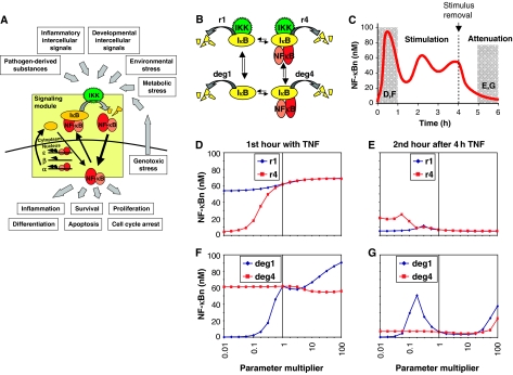
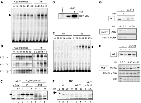
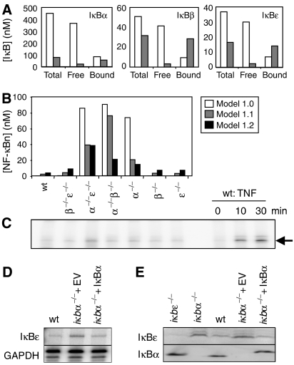
Cellular signal transduction pathways are usually studied following administration of an external stimulus. However, disease-associated aberrant activity of the pathway is often due to misregulation of the equilibrium state. The transcription factor NF-kappaB is typically described as being held inactive in the cytoplasm by binding its inhibitor, IkappaB, until an external stimulus triggers IkappaB degradation through an IkappaB kinase-dependent degradation pathway. Combining genetic, biochemical, and computational tools, we investigate steady-state regulation of the NF-kappaB signaling module and its impact on stimulus responsiveness. We present newly measured in vivo degradation rate constants for NF-kappaB-bound and -unbound IkappaB proteins that are critical for accurate computational predictions of steady-state IkappaB protein levels and basal NF-kappaB activity. Simulations reveal a homeostatic NF-kappaB signaling module in which differential degradation rates of free and bound pools of IkappaB represent a novel cross-regulation mechanism that imparts functional robustness to the signaling module.


Figures

Figure 1
Figure 1
Exploring the relative importance of IκB degradation mechanisms by computational parameter sensitivity analysis. (A) Schematic of the NF-κB signaling module and its physiological importance in the transduction of diverse inflammatory, developmental, and stress signals. (B) Illustration of the four IκB degradation pathways within the NF-κB signaling module. deg1 and deg4 are IKK-independent degradation rate constants for free and bound IκBα. r1 and r4 are IKK-dependent degradation rate constants for free and bound IκBα. (C) Computational simulation of NF-κB activation over a 6-h time course. TNF stimulation begins at time 0, and is removed at 4 h. Mean activity in the first hour of stimulation and the second hour after removal of the stimulus (shaded in gray) were used to create the plots in (D–F) and (G). (DG) Graphs showing the average nuclear NF-κB (y axis) during the first hour (D, F) or during the second hour after 4 h (E, G) of TNF stimulation for different values (x axis) of the IKK-dependent (D, E) or -independent (F, G) degradation rate constants of free (blue line) and bound (red line) IκB.
Figure 2
Figure 2
Experimental studies of degradation pathways of NF-κB-bound and -free IκB proteins. (A) NF-κB activity as measured by EMSA of nuclear extracts from wild-type cells treated with 10 μg/ml CHX or 1 ng/ml TNF for indicated times. (B) NF-κB activity as measured by EMSA of nuclear extracts from ikbβ−/−ɛ−/−, ikbα−/−ɛ−/−, or ikbα−/−β/− cells treated with 10 μg/ml CHX or 1 ng/ml TNF. (C) Western blot for IκBα in CHX-treated wild-type and nfkb−/− cells. The first two lanes show iκbα−/−β−/−ɛ−/− extract and iκbα−/−β−/−ɛ−/− extract mixed with 5% wild-type extract to show the protein level of IκBα in the nfkb−/− cells was approximately 5% that in the wild-type cells at time zero. (D) Cytoplasmic extracts of wild-type cells were immunoprecipitated with IKKγ antibody and subject to an in vitro kinase assay. In the ‘mock' lane, no antibody was added during the IP. (E) NF-κB activity as measured by EMSA of nuclear extracts from ikkα−/−β−/− or wild-type MEFs treated with 10 μg/ml CHX. (F) Western blot for IκBα of protein extracts from TNF (1 ng/ml)-treated wild-type or rela−/−crel−/−nfkb1−/− cells. (G) Western blots for IκBα of protein extracts from TNF-treated wild-type cells (top panel) in the presence or absence of the IKK-inhibitor sc-514. Bottom panels show Western blots for IκBα of protein extracts from CHX (10 μg/ml)-treated cells in the presence or absence of sc-514. (H) Western blots for IκBα of protein extracts from wild-type cells treated with TNFα (1 ng/ml) with or without the presence of the proteasome inhibitor MG132 (top panel). Western blots for IκBα of protein extracts from nfkb−/− cells treated with 10 μg/ml CHX, 10 μM MG132, or both.
Figure 3
Figure 3
An improved model of the homeostatic NF-κB signaling module. (A) Model calculations of IκBα, β, and ɛ protein levels (nM) in unstimulated cells for total IκB, free IκB, or NF-κB-bound IκB. Model 1.0 predictions are white bars, model 1.1 predictions are gray bars. (B) Model calculations of NF-κB activity (nM) in unstimulated wild-type and ikb−/− cells predicted by model version 1.0 (white bars), version 1.1 (gray bars), and version 1.2 (black bars). (C) NF-κB activity in untreated cells as measured by EMSA of nuclear extracts from the iκb−/− cell genotype labeled above each lane. The last three lanes are controls for the NF-κB band and are nuclear extracts from wild-type cells treated with TNF (1 ng/ml). (D) RNase protection assay showing levels of IκBɛ mRNA in untreated wild-type cells, iκbα−/− cells with empty vector control, or iκbα−/− cells expressing a retroviral iκbα transgene. GAPDH is used as a loading control. (E) Western blots for IκBɛ and IκBα in resting cells. The cell genotype is listed above each lane.

Similar articles

Cited by

References

    1. Alvarez-Castelao B, Castano JG (2005) Mechanism of direct degradation of IkappaBalpha by 20S proteasome. FEBS Lett 579: 4797–4802 - PubMed
    1. Bren GD, Pennington KN, Paya CV (2000) PKC-zeta-associated CK2 participates in the turnover of free IkappaBalpha. J Mol Biol 297: 1245–1258 - PubMed
    1. Brivanlou AH, Darnell JE Jr (2002) Signal transduction and the control of gene expression. Science 295: 813–818 - PubMed
    1. Ghosh S, May MJ, Kopp EB (1998) NF-kappa B and Rel proteins: evolutionarily conserved mediators of immune responses. Annu Rev Immunol 16: 225–260 - PubMed
    1. Hoffmann A, Baltimore D (2006) Circuitry of nuclear factor kappaB signaling. Immunol Rev 210: 171–186 - PubMed

Publication types

MeSH terms