Review
. 2007 Aug;151(7):930-40.
doi: 10.1038/sj.bjp.0707272. Epub 2007 May 8.

Nitric oxide homeostasis as a target for drug additives to cardioplegia

Affiliations
Review

Nitric oxide homeostasis as a target for drug additives to cardioplegia

B K Podesser et al. Br J Pharmacol. 2007 Aug.

Abstract

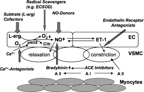
The vascular endothelium of the coronary arteries has been identified as the important organ that locally regulates coronary perfusion and cardiac function by paracrine secretion of nitric oxide (NO) and vasoactive peptides. NO is constitutively produced in endothelial cells by endothelial nitric oxide synthase (eNOS). NO derived from this enzyme exerts important biological functions including vasodilatation, scavenging of superoxide and inhibition of platelet aggregation. Routine cardiac surgery or cardiologic interventions lead to a serious temporary or persistent disturbance in NO homeostasis. The clinical consequences are "endothelial dysfunction", leading to "myocardial dysfunction": no- or low-reflow phenomenon and temporary reduction of myocardial pump function. Uncoupling of eNOS (one electron transfer to molecular oxygen, the second substrate of eNOS) during ischemia-reperfusion due to diminished availability of L-arginine and/or tetrahydrobiopterin is even discussed as one major source of superoxide formation. Therefore maintenance of normal NO homeostasis seems to be an important factor protecting from ischemia/reperfusion (I/R) injury. Both, the clinical situations of cardioplegic arrest as well as hypothermic cardioplegic storage are followed by reperfusion. However, the presently used cardioplegic solutions to arrest and/or store the heart, thereby reducing myocardial oxygen consumption and metabolism, are designed to preserve myocytes mainly and not endothelial cells. This review will focus on possible drug additives to cardioplegia, which may help to maintain normal NO homeostasis after I/R.


Figures

Figure 1
Figure 1
Nitric oxide (NO) homeostasis as a target for drug additives to cardioplegia – possible routes: following reperfusion after cardioplegia, the levels of NO are decreased mainly due to ‘uncoupling' of endothelial nitric oxide synthase (eNOS), which leads to enhanced level of superoxide (O2). This results in increased vascular tone due to reduced bioavailability of NO and therefore reduced relaxation as well as enhanced production of the vasoconstrictors endothelin-1 (ET-1) and angiotensin II (A II). This reperfusion injury can be attenuated by enhancing the levels of bioavailable NO with substrates/cofactors of eNOS, NO-donors (feedback inhibition and prevention of ‘uncoupling'), bradykinin, blocking ET-I receptors, blocking the receptors (AT-I) or production (ACE- inhibitors) of angiotensin II and free radical scavengers of superoxide such as extracellular superoxide dismutase (ECSOD). Calcium antagonists on the other hand can counteract the initial Ca2+ increase at the onset of ischemia and prevent eNOS stimulation. The enhanced (normalized) levels of bioavailable NO, for example, attenuate/prevent inflammation by inhibiting the activation of the proinflammatory transcription factor nuclear factor kappa B (NF-κB) and cell adhesion molecules. As consequence this results in a decreased expression of proinflammatory cytokines and reduced leukocyte (neutrophil) adhesion. The above figure mainly illustrates the possible routes of intervention.
Figure 2
Figure 2
S-nitroso human serum albumin (S-NO-HSA) attenuates ischemia/reperfusion injury after prolonged cardioplegic arrest in isolated rabbit hearts: in an isolated erythrocyte-perfused rabbit working heart model after assessment of hemodynamic baseline values hearts were randomly assigned to receive S-NO-HSA (0.2 μmol/100ml, n=8), L-arginine (10 mmol/100 ml, n=8) or albumin (control; 0.2 μmol/100 ml, n=8). After 20 min of infusion, the hearts were arrested and stored in Celsior solution (4°C) enriched with respective drugs for 6 h, followed by 75 min of reperfusion. Postischemic recovery of hemodynamic parameters are shown. (a) Time course of cardiac output; *P<0.05, **P<0.01 S-NO-HSA versus control. ΔP<0.05 S-NO-HSA versus L-arginine;P<0.05 L-arginine versus control. (b) Time course of coronary flow; *P<0.05, **P<0.01 S-NO-HSA versus control. ΔP<0.05 S-NO-HSA versus L-arginine. (c) Time course of left atrial pressure; *P<0.05 S-NO-HSA versus control. (d) Myocardial oxygen consumption; **P<0.01 S-NO-HSA versus control. ††P<0.01 L-arginine versus control. (This figure is from Semsroth et al., 2005: J Heart Lung Transplant 24: 2226–2234© Elsevier Publishing Group with permission.)

Similar articles

Cited by

References

    1. Ambrosio G, Flaherty JT, Duilio C, Tritto I, Santoro G, Elia PP, et al. Oxygen radicals generated at reflow induce peroxidation of membrane lipids in reperfused hearts. J Clin Invest. 1991;87:2056–2066. - PMC - PubMed
    1. Amrani M, Gray CC, Smolenski RT, Goodwin AT, London A, Yacoub MH. The effect of L-arginine on myocardial recovery after cardioplegic arrest and ischemia under moderate and deep hypothermia. Circulation. 1997;96 (Suppl 9):II274–II279. - PubMed
    1. Arai H, Hori S, Aramori I, Ohkubo H, Nakashini S. Cloning and expression of a cDNA encoding an endothelin receptor. Nature. 1990;3458:730–732. - PubMed
    1. Azuma H, Ishikawa M, Sekizaki S. Endothelium–dependent inhibition of platelet aggregation. Br J Pharmacol. 1986;88:411–415. - PMC - PubMed
    1. Balligand JL, Cannon PJ. Nitric oxide synthases and cardiac muscle. Arterioscler Thromb Vasc Biol. 1997;17:1846–1858. - PubMed

Publication types

MeSH terms