. 2006 Oct-Dec;1(4):e1-11.
doi: 10.4161/epi.1.4.3514.

Reduced MeCP2 expression is frequent in autism frontal cortex and correlates with aberrant MECP2 promoter methylation

Affiliations

Reduced MeCP2 expression is frequent in autism frontal cortex and correlates with aberrant MECP2 promoter methylation

Raman P Nagarajan et al. Epigenetics. 2006 Oct-Dec.

Abstract

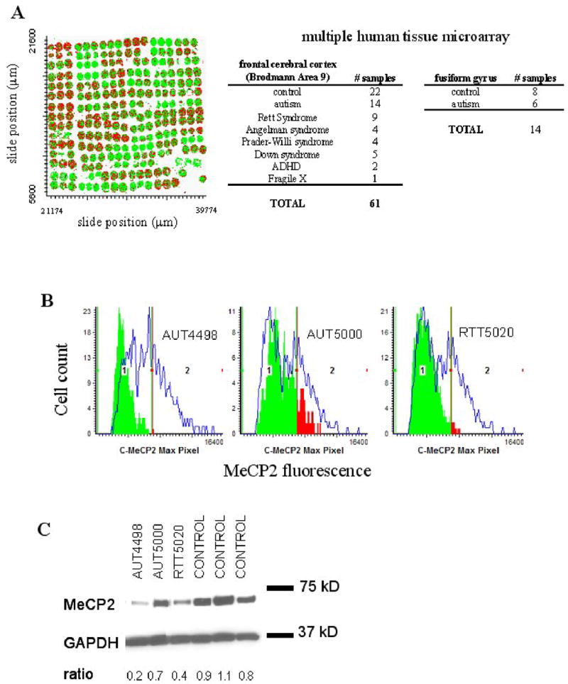
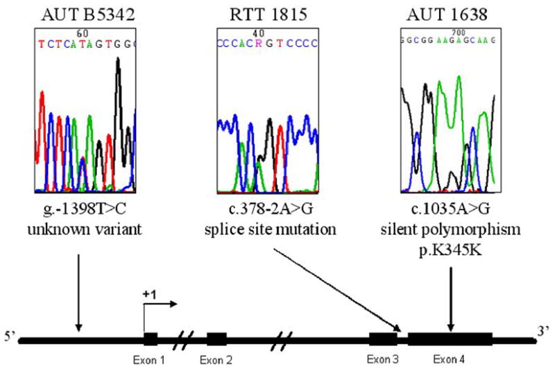
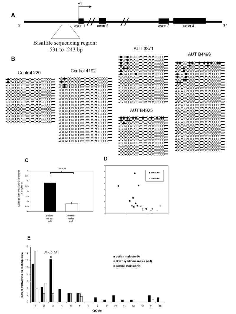
Mutations in MECP2, encoding methyl CpG binding protein 2 (MeCP2), cause most cases of Rett syndrome (RTT), an X-linked neurodevelopmental disorder. Both RTT and autism are "pervasive developmental disorders" and share a loss of social, cognitive and language skills and a gain in repetitive stereotyped behavior, following apparently normal perinatal development. Although MECP2 coding mutations are a rare cause of autism, MeCP2 expression defects were previously found in autism brain. To further study the role of MeCP2 in autism spectrum disorders (ASDs), we determined the frequency of MeCP2 expression defects in brain samples from autism and other ASDs. We also tested the hypotheses that MECP2 promoter mutations or aberrant promoter methylation correlate with reduced expression in cases of idiopathic autism. MeCP2 immunofluorescence in autism and other neurodevelopmental disorders was quantified by laser scanning cytometry and compared with control postmortem cerebral cortex samples on a large tissue microarray. A significant reduction in MeCP2 expression compared to age-matched controls was found in 11/14 autism (79%), 9/9 RTT (100%), 4/4 Angelman syndrome (100%), 3/4 Prader-Willi syndrome (75%), 3/5 Down syndrome (60%), and 2/2 attention deficit hyperactivity disorder (100%) frontal cortex samples. One autism female was heterozygous for a rare MECP2 promoter variant that correlated with reduced MeCP2 expression. A more frequent occurrence was significantly increased MECP2 promoter methylation in autism male frontal cortex compared to controls. Furthermore, percent promoter methylation of MECP2 significantly correlated with reduced MeCP2 protein expression. These results suggest that both genetic and epigenetic defects lead to reduced MeCP2 expression and may be important in the complex etiology of autism.


Figures

Figure 1
Figure 1
Decreased MeCP2 protein expression in autism samples detected by IF/LSC and immunoblot. A. X,Y scattergram of multiple tissue microarray used for IF/LSC. Each colored circle is a 600 μm core of human cerebral cortex postmortem tissue, containing approximately 200–500 cell nuclei. Cell populations with low and high MeCP2 expression are differentiated by dividing the adult control sample MeCP2 histogram at the right half-max. All cells to the left of the gate are colored green (MeCP2lo), and all cells to the right of the gate are colored red (MeCP2hi) B. IF/LSC analysis of MeCP2 protein expression in three representative cerebral cortex samples. MeCP2 expression for autism and RTT samples are shown as green and red colored solid histograms. RTT 5020 and AUT 4498 show decreased MeCP2 protein expression compared to three closest age-matched controls (blue line overlay). AUT 5000 shows less significantly decreased MeCP2 protein expression compared to controls. C. Immunoblot analysis shows similar results as IF/LSC analysis in B. Anti-GAPDH was used as a loading control and MeCP2 expression is normalized to GAPDH and shown as a ratio below the blot image.
Figure 2
Figure 2
Sequencing of MECP2 coding and promoter sequences in brain genomic DNA. The 4 MECP2 coding exons, including intronexon boundaries, were sequenced in all neurodevelopmental disorder samples on the tissue microarray. For the MECP2 promoter, a 1.9 kb portion of the MECP2 promoter region was sequenced including 1630 bp upstream of the transcription start site, exon 1, and 254 bp of intron 1. For promoter sequencing, a total of 51.3 kb was analyzed (1.9 kb/27 individuals). Sequencing chromatograms for three brain samples with sequence variations (AUT B5342, RTT 1815, and AUT 1638) are shown above a schematic diagram of the MECP2 genomic region. Exons are indicated with filled rectangles and the transcription start site is shown as +1. Arrows underneath each sequencing chromatogram point to the approximate location of the sequence variant.
Figure 3
Figure 3
Bisulfite sequencing analysis of MECP2 promoter methylation in frontal cortex DNA from autism and control postmortem brain samples. A. Schematic diagram of the MECP2 genomic region showing exons (filled rectangles), the transcription start site (+1), and the bisulfite sequencing region (bracketed sequence). B. Representative bisulfite sequencing data from two control males (229 and 4192) and three autism males (AUT 3871, AUT B4925, and AUT B4498). Each line with circles represents an individual clone. Filled circles indicate methylated CpG sites, and open circles indicate unmethylated CpG sites. A minimum of 10 clones were analyzed for each brain sample. All bisulfite data are shown in Supplementary Figure I. C. Percent MECP2 methylation for autism (n=9), and control (n=9) males was calculated by dividing the number of methylated CpG sites by the total number of CpG sites assayed for each brain sample. Results are graphed as mean ± SEM, and significance was determined by t-test. A statistically significant difference between autism and controls was also observed by analyzing the percentage of clones with one or more methylated sites out of the total number of clones for each sample (data not shown) D. Scatterplot showing percent MECP2 methylation and normalized MeCP2 expression for each brain sample. Autism samples are shown as filled circles and controls are shown as open squares. Regression was calculated for autism and controls together (R2=0.3235, P<0.05), as well as for autism (R2=0.1334) and control(R2=0.1065) samples alone. E. The percent methylation for each CpG site was compared between autism (n=9), Down syndrome (n=4), and control (n=9) males. CpG sites are shown on the x axis as in B. CpG site #3 showed a significant increase (P=0.0031, t-test) in autism vs. controls.

References

    1. Kriaucionis S, Bird A. DNA methylation and Rett syndrome. Hum Mol Genet. 2003;12:R221–227. Spec No 2. - PubMed
    1. Nicolson R, Szatmari P. Genetic neurodevelopmental influences in autistic disorder. Can J Psychiatry. 2003;48:526–537. - PubMed
    1. Lamb JA, Moore J, Bailey A, Monaco AP. Autism: recent molecular genetic advances. Hum Mol Genet. 2000;9:861–868. - PubMed
    1. Tager-Flusberg H, Joseph R, Folstein S. Current directions in research on autism. Ment Retard Dev Disabil Res Rev. 2001;7:21–29. - PubMed
    1. Amir RE, Van den Veyver IB, Wan M, Tran CQ, Francke U, Zoghbi HY. Rett syndrome is caused by mutations in X-linked MECP2 encoding methyl-CpG-binding protein 2. Nat Genet. 1999;23:185–188. - PubMed

Publication types

Substances

LinkOut - more resources