. 2007 Aug;36(2):242-50.
doi: 10.1002/mus.20817.

Impact of gravity loading on post-stroke reaching and its relationship to weakness

Affiliations

Impact of gravity loading on post-stroke reaching and its relationship to weakness

Randall F Beer et al. Muscle Nerve. 2007 Aug.

Abstract

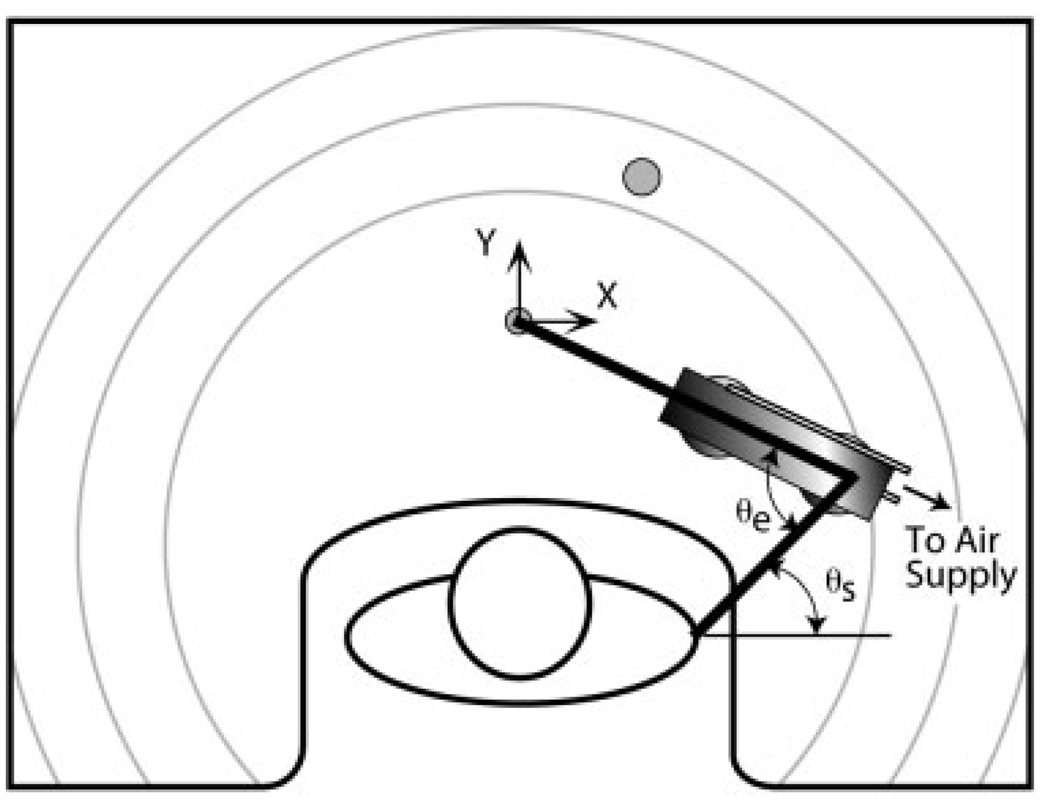
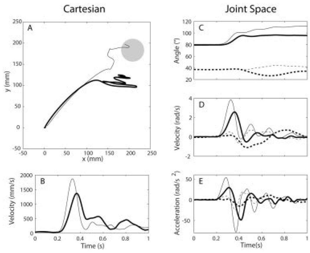
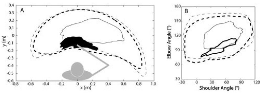
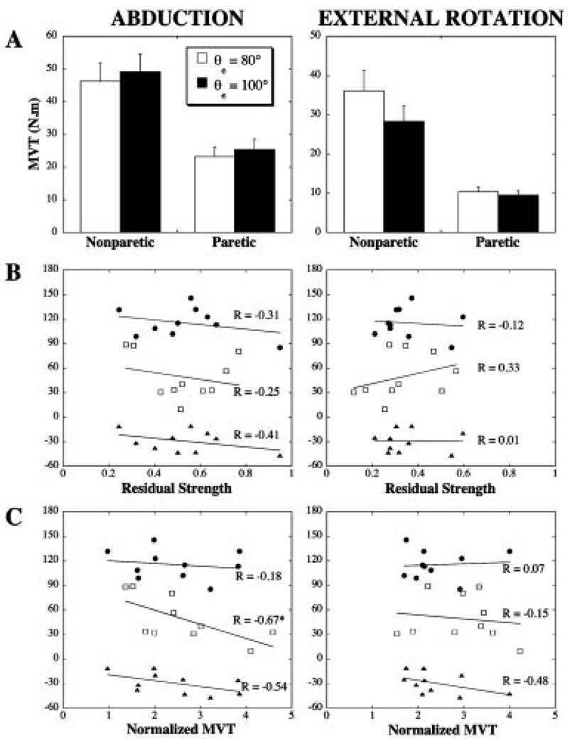
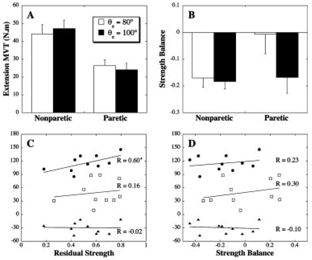
The ability to extend the elbow following stroke depends on the magnitude and direction of torques acting at the shoulder. The mechanisms underlying this link remain unclear. The purpose of this study was to evaluate whether the effects of shoulder loading on elbow function were related to weakness or its distribution in the paretic limb. Ten subjects with longstanding hemiparesis performed movements with the arm either passively supported against gravity by an air bearing, or by activation of shoulder muscles. Isometric maximum voluntary torques at the elbow and shoulder were measured using a load cell. The speed and range of elbow extension movements were negatively impacted by actively supporting the paretic limb against gravity. However, the effects of gravity loading were not related to proximal weakness or abnormalities in the elbow flexor-extensor strength balance. The findings support the existence of abnormal descending motor commands that constrain the ability of stroke survivors to generate elbow extension torque in combination with abduction torque at the shoulder.


Figures

FIGURE 1
FIGURE 1
Schematic of the experimental arrangement (viewed from above). Movements were performed with the forearm in a horizontal plane and the arm abducted to 75°. Subjects completed point-to-point and active range of motion protocols, with the arm either passively supported on a lightweight air-bearing device or actively supported by the shoulder musculature. The target (shaded circle) for point-to-point movements was located to require a 30° extension rotation at the elbow, without change in the elbow Cartesian position. The active range of motion protocol required subjects to trace out the largest possible area in front of their body. Circular arcs on the table surface were used to encourage achievement of maximum reaching distance. The positions of the hand, elbow, and shoulder were measured in Cartesian coordinates using an optoelectric system and converted off-line into the corresponding shoulder horizontal flexion/extension (θs) and elbow (θe) angles.
FIGURE 2
FIGURE 2
Cartesian and joint kinematics for actively (thick lines) and passively (thin lines) supported point-to-point movements of the paretic limb. Mean hand paths and tangential velocity profiles are shown in (A) and (B), respectively. Mean joint angle, angular velocity, and angular acceleration profiles are shown for the elbow (solid lines) and shoulder (dashed lines) in (C–E). Trials were aligned at movement onset (at 0.2 s in (B–E)) for averaging. Positive ordinates correspond to elbow extension and shoulder horizontal flexion. Data is for subject 5 (see Table 1).
FIGURE 3
FIGURE 3
Typical results for the range of motion protocol for the paretic (solid lines) and nonparetic (dotted lines) limbs. (A) Work area of the hand with active (thick lines) and passive (thin lines) support of the arm against gravity. The work area of the paretic limb with active support is shaded for clarity. Underlying shoulder and elbow motions are shown in (B), with a 180° elbow angle representing a straight limb and positive values of shoulder angle indicating a flexed position of the upper arm segment in the horizontal plane. The plotted data represent envelopes derived from 10 trials performed by subject 7.
FIGURE 4
FIGURE 4
Proximal weakness and its relationship to deficits in actively supported elbow extension. (A) Maximum voluntary abduction and external rotation torques (group mean with SEM). (B) Correlational analyses between movement parameters and residual strength. (C) Correlational analyses between movement parameters and normalized abduction and external rotation MVTs (quotient of the MVT and the corresponding torque required to support the arm against gravity). *Significant correlation, P< 0.05. ● Extension AROM, active support condition (°); □ normalized peak elbow acceleration (%); ▲ change in AROM with active support (°).
FIGURE 5
FIGURE 5
(A) Elbow extension MVT (group mean with SEM). (B) Elbow strength balance (SB) (group mean with SEM). A positive SB represents a larger MVT in extension, compared to flexion. Correlational analyses between movement parameters and elbow extension residual strength (C) and elbow strength balance (D)+Marginally significant correlation, P= 0.07. ● Extension AROM, active support condition (°); □ normalized peak elbow acceleration (%); ▲ change in AROM with active support (°).

References

    1. Barbeau H, Visintin M. Optimal outcomes obtained with body-weight support combined with treadmill training in stroke subjects. Arch Phys Med Rehabil. 2003;84:1458–1465. - PubMed
    1. Beer RF, Dewald JP, Dawson ML, Rymer WZ. Target-dependent differences between free and constrained arm movements in chronic hemiparesis. Exp Brain Res. 2004;156:458–470. - PubMed
    1. Beer RF, Dewald JP, Rymer WZ. Deficits in the coordination of multijoint arm movements in patients with hemiparesis: evidence for disturbed control of limb dynamics. Exp Brain Res. 2000;131:305–319. - PubMed
    1. Beer RF, Given JD, Dewald JP. Task-dependent weakness at the elbow in patients with hemiparesis. Arch Phys Med Rehabil. 1999;80:766–772. - PubMed
    1. Bohannon RW, Smith MB. Interrater reliability of a modified Ashworth scale of muscle spasticity. Phys Ther. 1987;67:206–207. - PubMed

Publication types