. 1991 Dec 15;266(35):23945-52.

The carboxyl terminus of the smooth muscle myosin light chain kinase is expressed as an independent protein, telokin

Affiliations

The carboxyl terminus of the smooth muscle myosin light chain kinase is expressed as an independent protein, telokin

P J Gallagher et al. J Biol Chem. .

Abstract

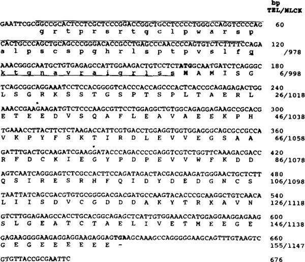
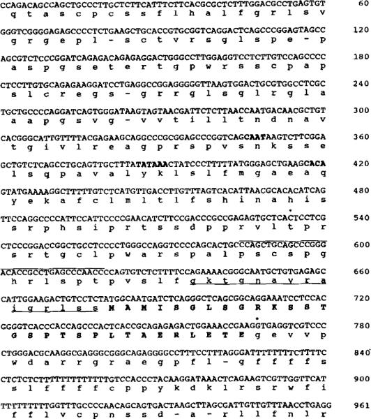
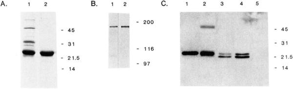
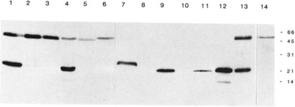
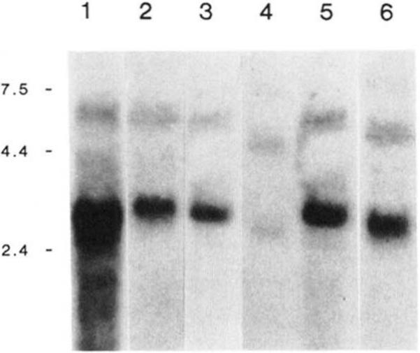
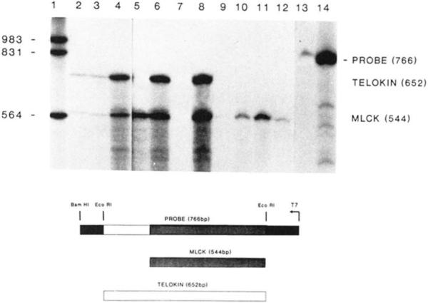
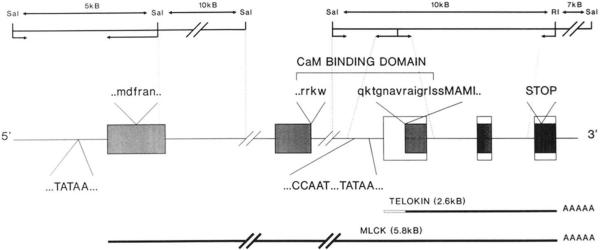
It has been proposed that the carboxyl terminus of the smooth muscle myosin light chain kinase is expressed as an independent protein. This protein has been purified from tissues and named telokin (Ito, M., Dabrowska, R., Guerriero, V., Jr., and Hartshorne, D. J. (1989) J. Biol. Chem. 264, 13971-13974). In this study we have isolated and characterized cDNA and genomic clones encoding telokin. Analysis of a genomic DNA clone suggests that the mRNA encoding telokin arises from a promoter which appears to be located within an intron of the smooth muscle myosin light chain kinase (MLCK) gene. This intron interrupts exons encoding the calmodulin binding domain of the kinase. The amino acid sequence deduced from the cDNA predicts that telokin is identical to the carboxyl-terminal 155 residues of the smooth muscle MLCK. Unlike the smooth muscle MLCK which is expressed in both smooth and non-muscle tissues, telokin is expressed in some smooth muscle tissues but has not been detected in aortic smooth muscle or in any non-muscle tissues.


Figures

Fig. 1
Fig. 1. Nucleotide and deduced amino acid sequence of the cDNA encoding the rabbit uterine telokin
The nucleotide sequence of the 676-bp cDNA clone identified in an MLCK-specific, oligonucleotide-primed library is shown. The 5′-noncoding region has been translated and is shown in lowercase letters. Nucleotides that are overlined (bp 9–116) are unique to the telokin cDNA; the remainder of the sequence is identical to that present in the rabbit smooth muscle MLCK (bp 3237–3787) (Gallagher et al., 1991), excluding the linkers (bp 1–8, 668–676). Residues that are underlined are within the coding region of the rabbit uterine smooth muscle MLCK. The nucleotides for translational initiation and termination are shown in boldface. An asterisk indicates the positions of introns which have been identified from sequence of the telokin gene. Numbers on the right side correspond to nucleotide sequence of the telokin cDNA (above) and amino acids of rabbit telokin/smooth muscle MLCK cDNAs (below).
Fig. 2
Fig. 2. Nucleotide sequence of a portion of the rabbit telokin gene
The nucleotide and deduced amino acid sequence of the rabbit telokin gene are shown. Residues shown in boldface capital letters are included in the coding region of telokin and the smooth muscle MLCK. Residues in lowercase letters and underlined are within the coding region of the rabbit smooth muscle MLCK and are within the predicted 5′-noncoding region in the telokin cDNA. An asterisk indicates the position of an intron in the rabbit telokin gene. Nucleotides that are overlined correspond to a primer used in the primer extension analysis. Nucleotides that are in boldface are those that are proposed as comprising the TATA box and transcriptional start site for the 2.6-kb mRNA encoding telokin.
Fig. 3
Fig. 3. Characterization of anti-telokin polyclonal antibodies
Panel A, Coomassie stained, SDS-polyacrylamide gel electrophoresis (15% acrylamide gel) of the bacterial expressed and purified telokin. Lane 1, bacterial lysate of E. coli HMS 174(DE3)-pET3A-TEL; lane 2, purified telokin protein. Panel B, immunoblot analysis with anti-telokin antibodies to detect the 152-kDa MLCK in a transfected COS cell extract expressing rabbit smooth muscle MLCK (lane 1) and rabbit uterine tissue (lane 2). Panel C, immunoblot analysis with anti-telokin polyclonal antibodies to detect rabbit telokin protein in extracts prepared from COS cells transfected with CMV5-TEL. Lane 1, purified, bacterial expressed telokin, 1 ng; lane 2, rabbit uterine tissue (19 μg of total protein); lanes 3 and 4, 5 and 10 μl of a COS cell extract expressing CMV5-Tel; lane 5, 10 μl of a COS cell extract expressing rabbit smooth muscle MLCK
Fig. 4
Fig. 4. Immunoblot analysis of smooth and non-muscle rabbit, chicken, and bovine tissues
Immunoblot analysis of tissue extracts using polyclonal anti-telokin antibodies. The amount of total protein loaded per lane varies as indicated and is not representative of the relative amounts of immunoreactive protein present in these tissues. The following tissues were used to prepare extracts: lanes 1 and 13, rabbit uterus (20 μg); lane 2, rabbit trachea (89 μg); lane 3, rabbit aorta (86 μg); lane 4, rabbit ileum (42 μg); lane 5, rabbit kidney (84 μg); lane 6, rabbit adrenal gland (110 μg); lane 7, chicken gizzard (60 μg); lane 8, chicken liver (76 μg); lane 9, bovine trachea (44 μg); lane 10, bovine adrenal gland (264 μg); lanes 11 and 12, purified telokin (1 and 5 ng, respectively); lane 14 represents an immunoblot performed using preimmune antiserum and 20 μg of rabbit uterine tissue.
Fig. 5
Fig. 5. Northern analysis of RNA isolated from smooth muscle tissues
Panel A, Northern analysis of RNA isolated from rabbit uteri (lane 1, 15 μg), rabbit ileum (lane 2, 20 μg), chicken gizzard (lane 3, 20 μg), human myometrial (lane 4, 20 μg), bovine trachea (lane 5, 30 μg), rat uteri (lane 6, 20 μg). The blot was probed with a 32P-labeled antisense RNA probe corresponding to the telokin cDNA. The positions of the RNA molecular weight standards are shown in kb on the left side of the figure. The lane representing rabbit uterine tissue was exposed for 24 h, and the other lanes were exposed for 72–96 h.
Fig. 6
Fig. 6. Ribonuclease protection analysis of rabbit smooth and non-muscle RNA
Hybridization of total RNA to the 32P-labeled antisense RNA was performed in solution (37 °C), and unprotected probe was digested with RNase as described under “Experimental Procedures.” Fragments that were protected by mRNAs present in the tissue were separated by electrophoresis on denaturing polyacrylamide gels and visualized by autoradiography. Lanes 2, 3, and 4, 2, 10 and 50 μg of rabbit uterine RNA, respectively; lane 5, 50 μg of rabbit aorta RNA; lanes 6 and 8, 10 and 50 μg of rabbit ileum RNA, respectively, lane 7 is blank; lanes 9, 10, and 11, 10, 50 and 100 μg of rabbit liver RNA, respectively; lane 12, 100 μg of rabbit kidney RNA; lanes 13 and 14, undigested probe. The sizes of end-labeled DNA molecular weight fragments (lane 1) are shown in bp on the left side of the figure. Indicated to the right side of the figure are the positions of the undigested probe, telokin, and MLCK fragments. A schematic below the figure illustrates the two protected fragments derived from the 766-bp riboprobe. The lightly shaded box represents the fragment protected by MLCK mRNA, and the open box represents the fragment protected by telokin mRNA.
Fig. 7
Fig. 7. Schematic representation of the rabbit smooth muscle MLCK gene
This schematic illustrates the proposed gene organization of the rabbit smooth muscle MLCK gene. The proposed structure has been based upon sequence obtained from portions of the rabbit MLCK gene and from cDNAs encoding the rabbit MLCK, and telokin and primer extension data. The upper portion of the figure shows a partial map of the two genomic DNA clones which have been characterized for this proposal. Portions of the gene which have been sequence are indicated by dotted lines. The genomic DNA comprising the extreme 5′ region of the gene has been described previously (Gallagher et al., 1991). The relative positions of the transcriptional start sites for the 5.8- and 2.6-kb mRNAs are indicated by TATAA. MLCK exons are represented by shaded boxes (not all exons are indicated), and telokin exons are represented by open boxes. Part of the amino acid sequence within the calmodulin (CaM) binding domain is shown above the linear schematic; residues indicated by lowercase letters are those present in MLCK, and residues in capital letters represent sequence at the translational start site for telokin.

References

    1. Adelstein RS, Conti MA, Daniel JL, Anderson W., Jr. Biochemistry and Pharmacology of Platelets. Elsevier Science Publishing Co.; North Holland, The Netherlands: 1975. pp. 101–119.
    1. Adelstein RS, Conti MA, Hathaway DR, Klee CB. J. Biol. Chem. 1978;253:8347–8350. - PubMed
    1. Andersson S, Davis DL, Dahlbäch H, Jörnvall H, Russell DW. J. Biol. Chem. 1989;264:8222–8229. - PubMed
    1. Arfin SM, Bradshaw RA. Biochemistry. 1988;27:7979–7984. - PubMed
    1. Bagchi IC, Kemp BE, Means AR. J. Biol. Chem. 1989;264:15843–15849. - PubMed

Publication types

MeSH terms

LinkOut - more resources