. 2007 Dec;56(12):1885-95.
doi: 10.1007/s00262-007-0331-2. Epub 2007 May 9.

HAGE, a cancer/testis antigen with potential for melanoma immunotherapy: identification of several MHC class I/II HAGE-derived immunogenic peptides

Affiliations

HAGE, a cancer/testis antigen with potential for melanoma immunotherapy: identification of several MHC class I/II HAGE-derived immunogenic peptides

Morgan G Mathieu et al. Cancer Immunol Immunother. 2007 Dec.

Abstract

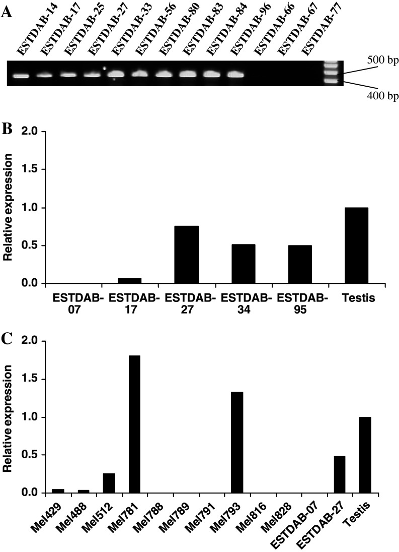
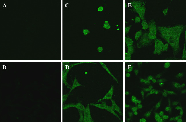
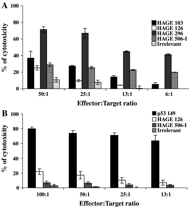
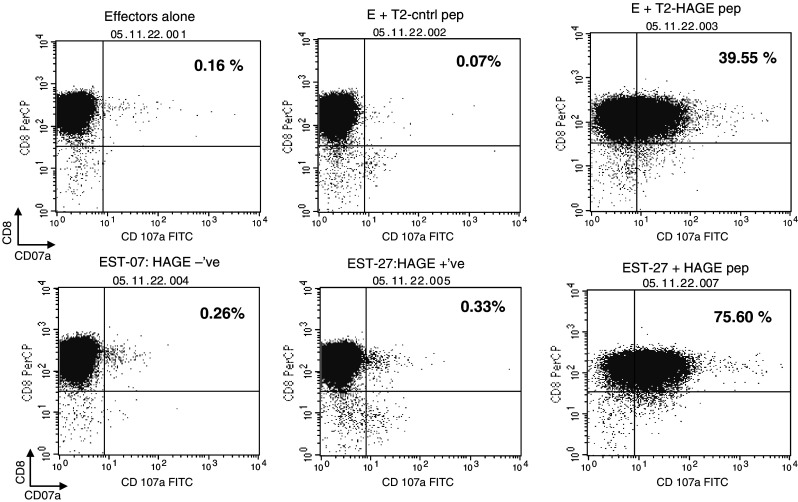
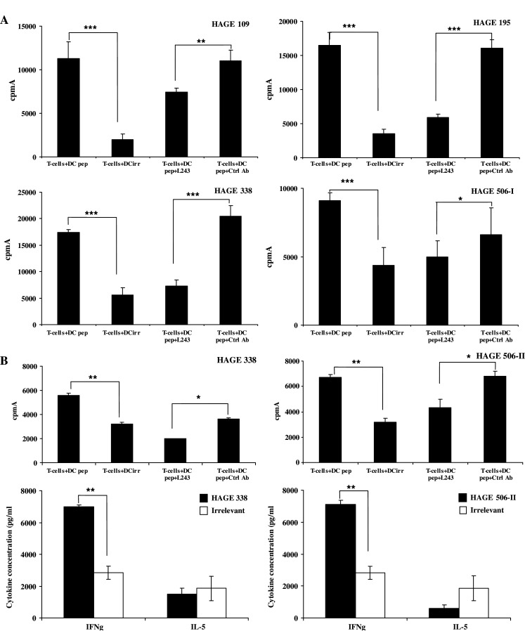
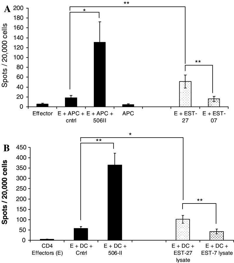
There remains a need to identify novel epitopes of potential tumour target antigens for use in immunotherapy of cancer. Here, several melanoma tissues and cell lines but not normal tissues were found to overexpress the cancer-testis antigen HAGE at the mRNA and protein level. We identified a HAGE-derived 15-mer peptide containing a shorter predicted MHC class I-binding sequence within a class II-binding sequence. However, only the longer peptide was found to be both endogenously processed and immunogenic for T cells in transgenic mice in vivo, as well as for human T cells in vitro. A different class I-binding peptide, not contained within a longer class II sequence, was subsequently found to be both immunogenic and endogenously processed in transgenic mice, as was a second class II epitope. These novel HAGE-derived epitopes may contribute to the range of immunotherapeutic targets for use in cancer vaccination programs.


Figures

Fig. 1
Fig. 1
Analysis of HAGE expression. a RT-PCR analysis of the expression of HAGE in ESTDAB melanoma cell lines using water as negative and testis as positive controls. PCR products for 18S ribosome and HAGE should be of 110 and 432 bp, respectively. Relative expression of HAGE obtained by real time RT-PCR analysis of b five tumour cell lines and c ten primary tumours from melanoma patients. The data are expressed relative to the mRNA level in normal testis, arbitrarily set as 1. The value of each sample was determined in triplicate reactions
Fig. 2
Fig. 2
HAGE protein expression in ESTDAB melanoma cell lines. Cells were grown onto chamber slides until 70% confluent was reached. Cells were then fixed and permeabilised and HAGE protein expression was assessed after a two step antibody staining. Immunofluorescence was observed under a confocal microscope (×40 magnification) in HAGE-positive ESTDAB-17 (c), -27 (d), -34 (e) and -95 (f) but not in HAGE-negative ESTDAB-07 (b). Cytoplasmic HAGE protein localisation can be observed. No non-specific secondary antibody staining was observed (a)
Fig. 3
Fig. 3
Immunogenicity and endogenous processing of class I HAGE derived peptides after peptides or DNA immunisation of HHDII mice and in vitro re-stimulation. Cytolytic properties of CTLs generated after immunisation of HHDII mice with class I HAGE derived peptides in combination with the HepB class II peptide in IFA are shown in panel a. Endogenous processing of any of these peptides was assessed after immunisation of HHDII with full length cDNA of either p53 as a control or HAGE, and one in vitro re-stimulation with p53 149 peptide or HAGE 506-I peptide, respectively, panel b. Target cells used were T2 pulsed with either p53 149, HAGE 506 class I (specific) or PAP135 (non-specific). The results presented here are the average of cytotoxicity obtained from six different mice for each peptide
Fig. 4
Fig. 4
HAGE 506-I-specific T cell clone co-cultured with HAGE-expressing melanoma cell line. A HAGE 506-I-specific CD8 T cell clone was challenged with either T2 cells pulsed with the HAGE 506-I or control peptide, or HAGE expressing (ESTDAB-27) or non-expressing (ESTDAB-07) HLA-A*0201-positive melanoma cell lines followed by CD107a measurement as a marker of cytolytic activity. HAGE expressing ESTDAB-27 was pulsed with the HAGE 506-I peptide as a positive control
Fig. 5
Fig. 5
Immunogenicity and endogenous processing of HAGE 506 class II peptide in DR4-transgenic mice. CD4+ T cells from three groups of two DR4 mice immunised with class II HAGE derived peptides (a) or full length HAGE cDNA (b) were stimulated in vitro with HAGE derived class II peptides, and then co-cultured with peptide-pulsed BM-DC. Peptide specificity is shown by means of a 3H-thymidine incorporation proliferation assay. **P < 0.01, unpaired Student’s t-test
Fig. 6
Fig. 6
Endogenous processing of HAGE 506-II peptide by a HAGE-expressing melanoma cell line or by autologous DC co-cultured with a cell lysate from a HAGE expressing cell line. Panel a shows the results of a HAGE 506-II-specific T-cell line challenged with either peptide-pulsed autologous PBMC or HLA-DR4+ melanoma cell-lines that were HAGE positive or negative. IFN-γ secretion by the T-cell line was monitored by ELISpot; for which 1 × 104 T cells (Effector E) were co-cultured for 40 h in an anti-IFN-γ-coated plate with either PBMC pulsed with HAGE 506-II or a control DR4 binding peptide or the melanoma cell lines (EST-07/27). The number of spots represents means of triplicate wells. Panel b shows the results of a HAGE 506-II specific T-cell line challenged with autologous DC pulsed with relevant or control (Ctrl) peptides or autologous immature monocyte-derived DC loaded with tumour cell lysate and matured overnight with TNFα and CD40L. The numbers of spots indicated are means of triplicate wells with the standard deviations indicated. *P < 0.05, **P < 0.01, unpaired Student’s t-test

Similar articles

Cited by

References

    1. Adams SP, Sahota SS, Mijovic A, Czepulkowski B, Padua RA, Mufti GJ, Guinn BA. Frequent expression of HAGE in chronic myeloid leukemias. Leukaemia. 2002;16:2238–2242. doi: 10.1038/sj.leu.2402732. - DOI - PubMed
    1. Assudani DP, Horton RB, Mathieu MG, McArdle SE, Rees RC. The role of CD4+ T cell help in cancer immunity and the formulation of novel cancer vaccines. Cancer Immunol Immunother. 2006;56:70–80. doi: 10.1007/s00262-006-0154-6. - DOI - PMC - PubMed
    1. Cordin O, Banroques J, Tanner NK, Linder P. The DEAD-box protein family of RNA helicases. Gene. 2006;367:17–37. doi: 10.1016/j.gene.2005.10.019. - DOI - PubMed
    1. Disis ML, Gralow JR, Bernhard H, Hand SL, Rubin WD, Cheever MA. Peptide-based, but not whole protein, vaccines elicit immunity to HER-2/neu, oncogenic self-protein. J Immunol. 1996;156:3151–3158. - PubMed
    1. Dissanayake SK, Tuera N, Ostrand-Rosenberg S. Presentation of endogenously synthesized MHC class II-restricted epitopes by MHC class II cancer vaccines is independent of transporter associated with Ag processing and the proteasome. J Immunol. 2005;174:1811–1819. - PubMed

Publication types

MeSH terms