. 2007 Jun;6(6):2351-66.
doi: 10.1021/pr0701411. Epub 2007 May 9.

Characterization of molecular recognition features, MoRFs, and their binding partners

Affiliations

Characterization of molecular recognition features, MoRFs, and their binding partners

Vladimir Vacic et al. J Proteome Res. 2007 Jun.

Abstract

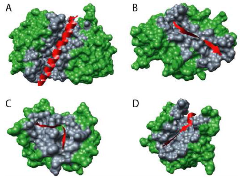
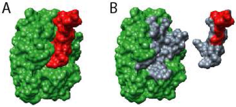
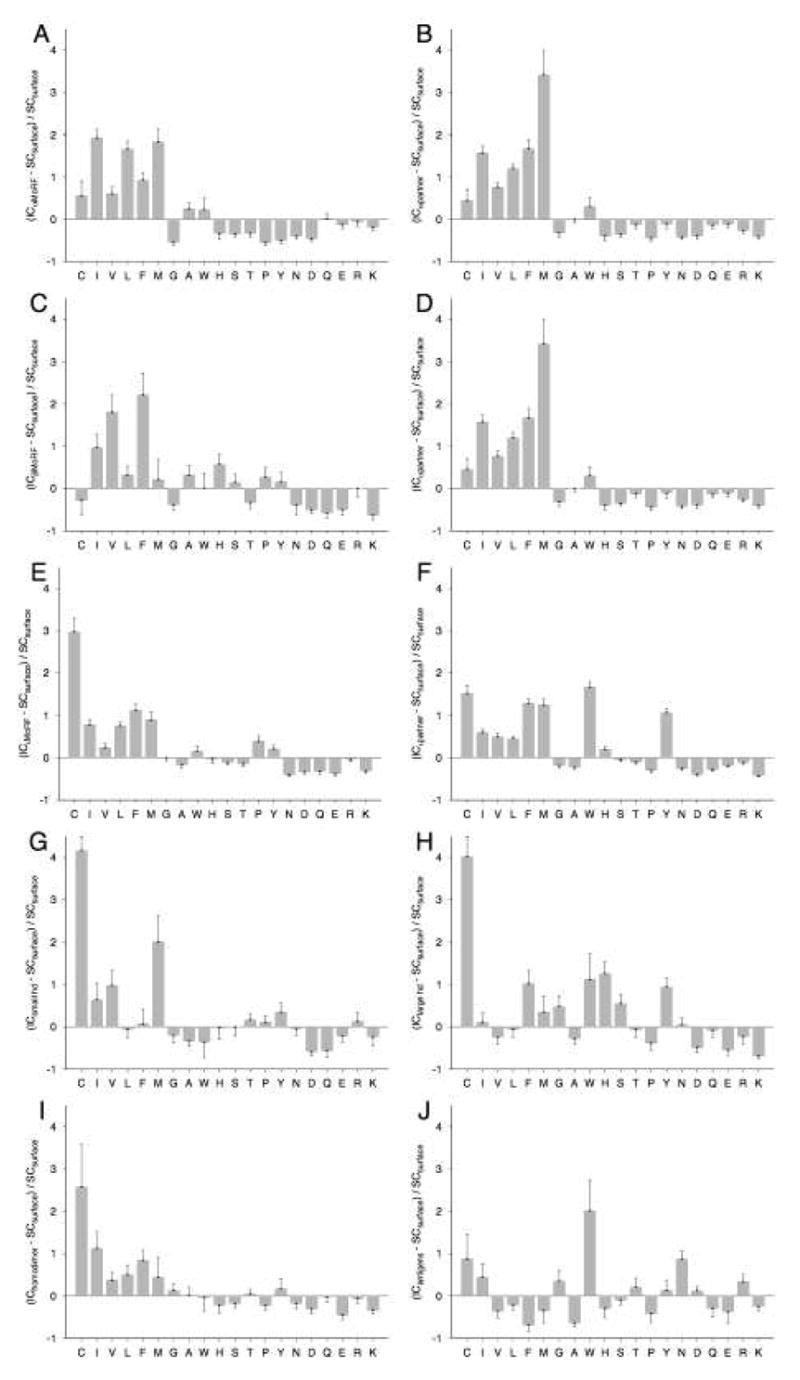
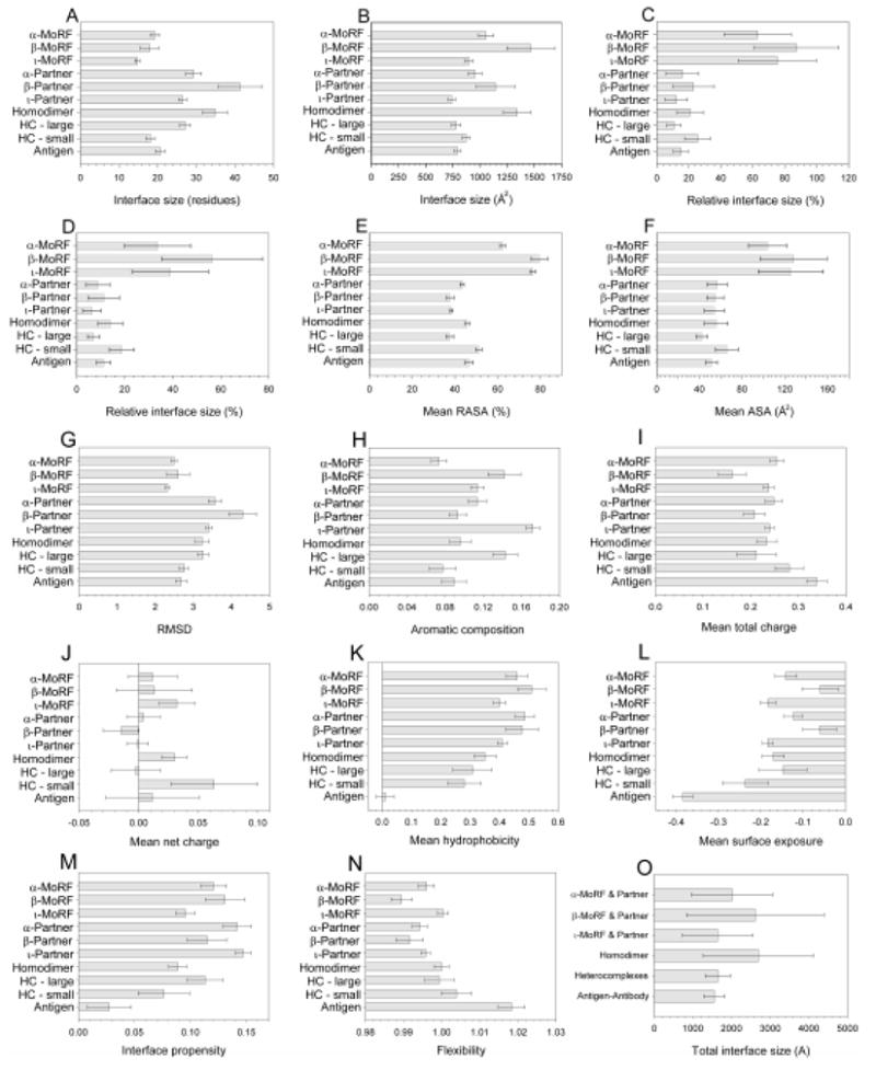
Molecular Recognition Features (MoRFs) are short, interaction-prone segments of protein disorder that undergo disorder-to-order transitions upon specific binding, representing a specific class of intrinsically disordered regions that exhibit molecular recognition and binding functions. MoRFs are common in various proteomes and occupy a unique structural and functional niche in which function is a direct consequence of intrinsic disorder. Example MoRFs collected from the Protein Data Bank (PDB) have been divided into three subtypes according to their structures in the bound state: alpha-MoRFs form alpha-helices, beta-MoRFs form beta-strands, and iota-MoRFs form structures without a regular pattern of backbone hydrogen bonds. These example MoRFs were indicated to be intrinsically disordered in the absence of their binding partners by several criteria. In this study, we used several geometric and physiochemical criteria to examine the properties of 62 alpha-, 20 beta-, and 176 iota-MoRF complex structures. Interface residues were examined by calculating differences in accessible surface area between the complex and isolated monomers. The compositions and physiochemical properties of MoRF and MoRF partner interface residues were compared to the interface residues of homodimers, heterodimers, and antigen-antibody complexes. Our analysis indicates that there are significant differences in residue composition and several geometric and physicochemical properties that can be used to discriminate, with a high degree of accuracy, between various interfaces in protein interaction data sets. Implications of these findings for the development of MoRF-partner interaction predictors are discussed. In addition, structural changes upon MoRF-to-partner complex formation were examined for several illustrative examples.


Figures

Figure 1
Figure 1. Examples of types of MoRFs
MoRFs (red ribbons) and partners (green surface) are shown (A) An α-MoRF, Proteinase Inhibitor IA3, bound to Proteinase A (PDB entry 1DP5). (B) A β-MoRF, viral protein pVIc, bound to Human Adenovirus 2 Proteinase (PDB entry 1AVP). (C) An ι-MoRF, Amphiphysin, bound to α-adaptin C (PDB entry 1KY7). (D) A complex-MoRF, β-amyloid precursor protein (βAPP), bound to the PTB domain of the neuron specific protein X11 (PDB entry 1X11). Partner interfaces (grey surface) are also indicated.
Figure 2
Figure 2. Illustration of interface ΔASA and identification of contact residues
Shown for the purpose of illustration is an ι-MoRF, Bowman-Birk type Trypsin Inhibitor, (A, red surface) bound to Trypsinogen (A, green surface) taken from PDB entry 1G9I. ASA is calculated for the complex (A) and the artificial monomers (B) separately to obtain ΔASA of complex formation. ASA can be attributed to individual residues, thereby allowing determination of residues involved in binding (B, grey surfaces).
Figure 3
Figure 3. Compositional profiles of the interface residues of MoRFs
The interface composition (IC) profiles of (A) α-MoRF, (B) α-MoRF partner, (C) β-MoRF, (D) β-MoRF partner, (E) ι-MoRF, (F) ι-MoRF partner, (G) small protomer from heterodimers, (H) large protomer from heterodimers, (I) homodimer, and (J) antigen interfaces are shown relative to surface residues of monomeric structures. Error bars give one standard deviation estimated by 100,000 bootstrap iterations. Amino acids are arranged in the order of increasing surface exposure, from the residues most buried in globular proteins on the left-hand side to the most exposed ones on the right-hand side .
Figure 4
Figure 4. Characterization of interface sizes and surface areas
Means and one standard deviation, estimated by 100,000 bootstrap iterations, are shown for each of the datasets for all metrics: (A) number of interface residues, (B) ASA of interface residues (C) proportion of residues involved in the interface, (D) proportion of ASA involved in the interface, (E) RASA of interface residues, (F) total ASA per residue, (G) planarity, (H) aromatic residue composition, (I) total and (J) net charge, (K) hydrophobicity, (L) surface exposure, (M) interface propensity, and (N) flexibility. In addition, surface buried on complex formation as mean ΔASA (O) are shown for all complex types.
Figure 5
Figure 5. Examples of disorder-to-order transitions in β- and ι-MoRFs
The structures of MoRFs (red ribbons) bound to their respective partners (blue ribbons) are shown. PONDR VL-XT predictions for the entire proteins in which these MoRFs are found are also shown, where the position of the MoRFs in these sequences are indicated (red boxes). (A) A β-MoRF example from p21 bound to PCNA (PDB entry 1AXC), where the position of the CDK inhibitor domain is indicated (green box). (B) An ι-MoRF example from Nup2p bound to karyopherin Kap60 (PDB entry 1UN0), where the positions of the FXF(G) repeat region (blue box) and the Ran binding domain (yellow box) are indicated.
Figure 6
Figure 6. Structural changes in partners
Ribbon representation MoRF partners shown unbound (blue ribbons) and bound (green ribbons) to MoRFs (red ribbons). (A) Small scale structural alterations in CheY induced by binding of the MoRF region of FliM (PDB entries: unbound - 1U8T and bound – 1F4V). (B) Large scale structural alterations in calmodulin induced by binding to the MoRF of GAD (PDB entries: unbound - 1CLL and bound - 1NWD). (C) Partial disorder-to-order transition in PCNA induced by binding to the MoRF of FEN-1 (PDB entries: unbound - 1RWZ and bound - 1RXZ). (D) Partial order-to-disorder transition in Bcl-xL induced by binding to the MoRF of Bim (PDB entries: unbound - 1PQ0 and bound - 1PQ1)

Similar articles

Cited by

References

    1. Argos P. An investigation of protein subunit and domain interfaces. Protein Eng. 1988;2:101–113. - PubMed
    1. Chothia C, Janin J. Principles of protein-protein recognition. Nature. 1975;256:705–708. - PubMed
    1. Tsai CJ, Lin SL, Wolfson HJ, Nussinov R. Studies of protein-protein interfaces: a statistical analysis of the hydrophobic effect. Protein Sci. 1997;6:53–64. - PMC - PubMed
    1. Janin J, Chothia C. The structure of protein-protein recognition sites. J Biol Chem. 1990;265:16027–16030. - PubMed
    1. Korn AP, Burnett RM. Distribution and complementarity of hydropathy in multisubunit proteins. Proteins. 1991;9:37–55. - PubMed

Publication types