

Antibodies against oxidized phospholipids in laboratory tests exploring lupus anti-coagulant activity
- PMID: 17488295
- PMCID: PMC1942029
- DOI: 10.1111/j.1365-2249.2007.03404.x
Antibodies against oxidized phospholipids in laboratory tests exploring lupus anti-coagulant activity
Abstract
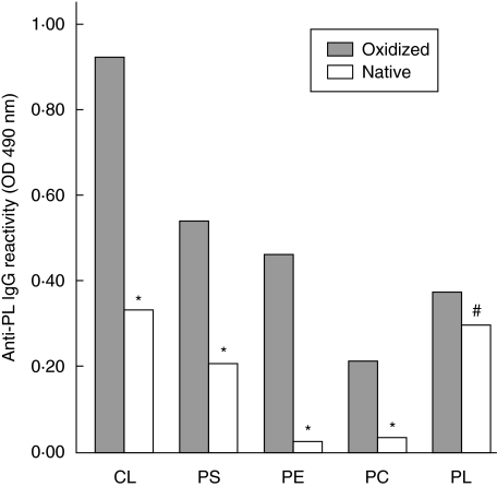
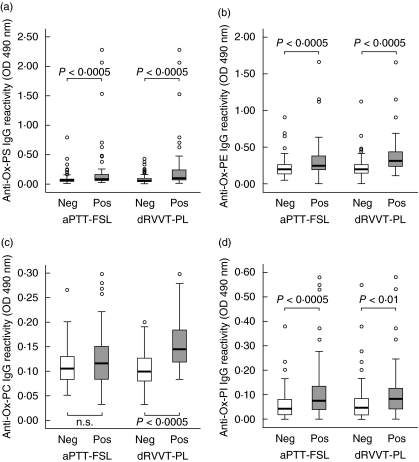
Lupus anti-coagulants (LA) are a variety of anti-phospholipid antibodies characterized by their capacity to interfere with phospholipid-dependent coagulation assays. LA are increasingly recognized as important predictors of thrombosis. However, the antigen specificity of LA is still poorly characterized. Growing evidence indicates that oxidized phospholipids are among the targets of anti-phospholipid antibodies. This prompted us to investigate the role of IgG directed against different oxidized phospholipids in 164 subjects without clotting factor defects that were tested for the presence of LA using a LA-sensitive activate partial thromboplastin time (aPTT-FSL) and a screening/confirmation assay based on diluted Russell's viper venom test (dRVVT-PL). The response to aPTT-FSL was significantly (P < 0.0005) associated with high titres of IgG against oxidized phosphatidylserine, phosphatidylethanolamine and phosphatidylinositol, whereas positivity to dRVVT-PL was associated with the elevation of IgG against oxidized phosphatidylserine, phosphatidylcholine, phosphatidylethanolamine (P < 0.0005) and phosphatidylinositol (P < 0.01). No difference in reactivity against oxidized cardiolipin was evident between the different groups. Positivity to the dRVVT-PL test was also associated significantly (P < 0.005) with the elevation of anti-cardiolipin and anti-beta(2)-glycoprotein-1 IgG. However, stepwise logistic regression demonstrated that IgG recognizing oxidized phosphatidylethanolamine and oxidized phosphatidylcholine were the only independent predictors of the response to dRVVT-PL assay, while IgG recognizing oxidized phosphatidylethanolamine and oxidized phosphatidylinositol were independent predictors of the response to aPTT-FSL test. In conclusion, autoantibodies against defined oxidized phospholipids are independent predictors of LA detection by aPTT-FSL or dRVVT-PL assays and might contribute to the variability often observed in the responses to the functional tests detecting LA.
Figures

Similar articles
-
IgG reactivity to phospholipid-bound beta(2)-glycoprotein I is the main determinant of the fraction of lupus anticoagulant activity quenched by addition of hexagonal (II) phase phospholipid in patients with the clinical suspicion of antiphospholipid-antibody syndrome.Haematologica. 1999 Sep;84(9):829-38. Haematologica. 1999. PMID: 10477458
-
Lupus anticoagulants and thrombosis: clinical association of different coagulation and immunologic tests.Thromb Haemost. 2000 Dec;84(6):1012-6. Thromb Haemost. 2000. PMID: 11154107
-
Utilization of dilute Russell's viper venom time to detect autoantibodies against beta 2-glycoprotein I which express anticoagulant activity in the presence but not in the absence of exogenous phospholipids.Thromb Haemost. 1997 Jan;77(1):123-6. Thromb Haemost. 1997. PMID: 9031461
-
The role of beta 2-glycoprotein I-dependent lupus anticoagulants in the pathogenesis of the antiphospholipid syndrome.Verh K Acad Geneeskd Belg. 2000;62(5):353-72. Verh K Acad Geneeskd Belg. 2000. PMID: 11144685 Review.
-
Use of the dilute Russell viper venom time (dRVVT): its importance and pitfalls.J Autoimmun. 2000 Sep;15(2):173-8. doi: 10.1006/jaut.2000.0414. J Autoimmun. 2000. PMID: 10968905 Review.
Cited by
-
Novel Autoimmune IgM Antibody Attenuates Atherosclerosis in IgM Deficient Low-Fat Diet-Fed, but Not Western Diet-Fed Apoe-/- Mice.Arterioscler Thromb Vasc Biol. 2020 Jan;40(1):206-219. doi: 10.1161/ATVBAHA.119.312771. Epub 2019 Oct 24. Arterioscler Thromb Vasc Biol. 2020. PMID: 31645128 Free PMC article.
-
Phospholipid-esterified eicosanoids are generated in agonist-activated human platelets and enhance tissue factor-dependent thrombin generation.J Biol Chem. 2010 Mar 5;285(10):6891-903. doi: 10.1074/jbc.M109.078428. Epub 2010 Jan 8. J Biol Chem. 2010. PMID: 20061396 Free PMC article.
References
-
- Bertolaccini ML, Hughes GR, Khamashta MA. Revisiting antiphospholipid antibodies: from targeting phospholipid to phospholipid binding proteins. Clin Lab. 2004;50:653–65. - PubMed
-
- Pierangeli SS, Harris EN. Clinical laboratory testing for antiphospholipid syndrome. Clin Chim Acta. 2005;357:17–23. - PubMed
-
- Levine JS, Branch DW, Rauch J. The antiphospholipid syndrom. N Engl J Med. 2003;346:752–63. - PubMed
-
- McNeil HP, Chesterman CN, Krilis SA. Immunological and clinical importance of antiphospholipid antibodies. Adv Immunol. 1991;49:193–280. - PubMed
-
- Veres K, Lakos G, Kerenyi A, et al. Antiphospholipid antibodies in acute coronary syndrome. Lupus. 2004;13:423–7. - PubMed
Publication types
MeSH terms
Substances
LinkOut - more resources
Full Text Sources
Other Literature Sources
