. 2007 Mar-Apr;11(2):286-98.
doi: 10.1111/j.1582-4934.2007.00023.x.

5'AMP-activated protein kinase alpha deficiency enhances stress-induced apoptosis in BHK and PC12 cells

Affiliations

5'AMP-activated protein kinase alpha deficiency enhances stress-induced apoptosis in BHK and PC12 cells

Margaret M Shaw et al. J Cell Mol Med. 2007 Mar-Apr.

Abstract

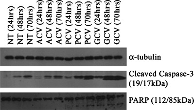
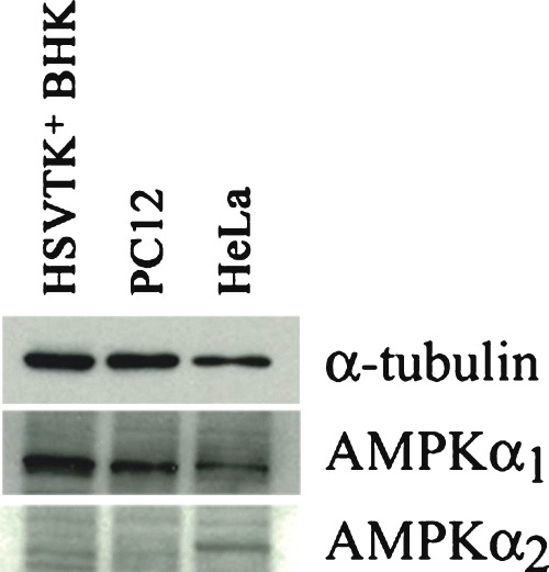
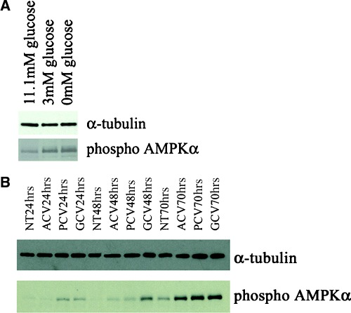
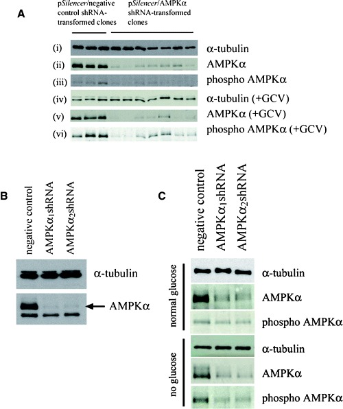
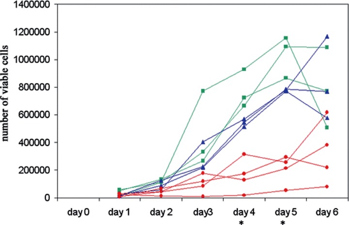
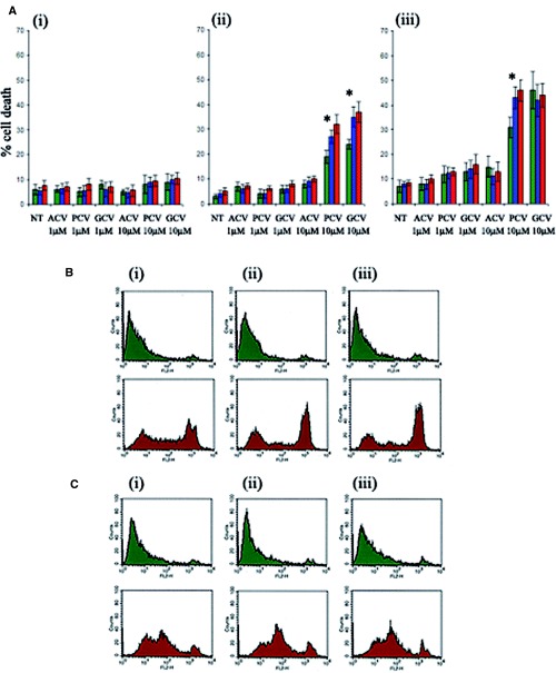
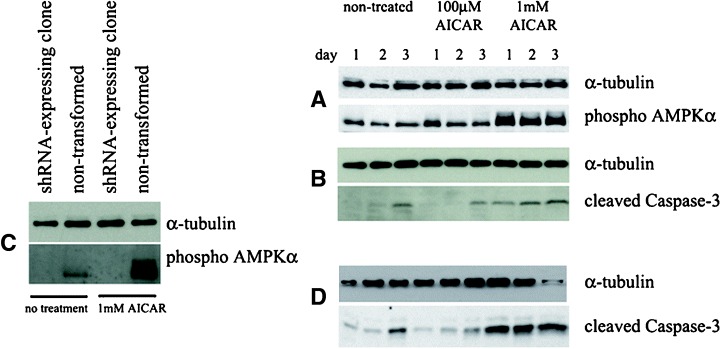
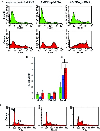
5'AMP-activated protein kinase (AMPK) activation occurs under a variety of stress conditions but the role of this enzyme in the promotion or inhibition of stress-induced cell death is unclear. To address this issue, we transformed two different cell lines with shRNA-expressing plasmids, targeting the alpha subunit of AMPK, and verified AMPKalpha downregulation. The cell lines were then stressed by exposure to medium without glucose (PC12 cells) or with the viral thymidine kinase-specific DNA replication inhibitors: acyclovir, penciclovir and ganciclovir (herpes simplex virus thymidine kinase-expressing Baby Hamster Kidney cells). In non-AMPK-downregulated cells, these stress treatments induced AMPK upregulation and phosphorylation, leaving open the question whether the association of AMPK activation with stress-induced cell death reflects a successful death-promoting or an ineffective death-inhibiting activity. In AMPKalpha-deficient cells (expressing AMPKalpha-specific shRNAs or treated with Compound C) exposure to low glucose medium or DNA replication inhibitors led to an enhancement of cell death, indicating that, under the conditions examined, the role of activated AMPK is not to promote, but to protect from or delay stress-induced cell death.


Figures

1
1
Apoptosis induction on treatment of HSVTK+ BHK cells with PCV and GCV. Western blot analysis shows high levels of cleaved Caspase-3 at 48 hrs and 70 hrs of treatment with PCV and GCV. PCV and GCV treatment also induces cleavage of the Caspase-3 substrate, PARP, which is not observed on treatment with ACV. (NT = non-treated control.)
2
2
Western blot showing AMPKα1/2 isoform expression in different cell types. Whereas both isoforms are detectable in HeLa cells, only AMPKα1 is found in HSVTK+ BHK and PC12 cells. Samples were equalized to α-tubulin (upper panel).
3
3
Western blot confirming enhanced phosphorylation of AMPKα in (A) PC12 cells exposed to low (3 mM) and glucose-free medium (11.1 mM glucose = normal culture conditions) and (B) HSVTK+ BHK cells treated with ACV, PCV or GCV.
4
4
(A) Western blot confirming reduction in levels of AMPKα in pSilencer-transformed HSVTK+ BHK cell clones, expressing the α1 or α2-targeting shRNA sequences (panel (ii)). Treatment with 10 μM GCV did not significantly increase total or phospho AMPKα expression in the AMPKα-downregulated clones (panels (v) and (vi)). Sample loadings in panels (ii) and (iii) were equalized to α-tubulin (panel (i)). Sample loadings in panels (v) and (vi) were equalized to α-tubulin (panel (iv) is a reprobe of the blot in panel (vi)). (B) Western blot confirming AMPKα downregulation in pSilencer/AMPKαshRNA-transformed HSVTK+ BHK cell populations, compared to the pSilencer/negative control shRNA-transformed population. (C) Western blot confirming AMPKα downregulation in pSilencer/AMPKαshRNA-transformed PC12 cells. Both total and phospho AMPKα remained downregulated during glucose deprivation. Sample loadings from the respective treatment groups were equalized to α-tubulin.
5
5
Growth curves of HSVTK+ BHK cell clones transformed with pSilencer/negative control shRNA (▪), pSilencer/AMPKα1shRNA (▴) or pSilencer/AMPKα2sh RNA (•), showing a slower growth rate in the pSilencer/ AMPKαshRNA-transformed clones, with most significant slowed growth in the α2 clones. (The differences between the individual groups (negative control, α1, α2) on days 4 and 5 are statistically significant, as measured by two-tail t-test, assuming equal variance (P < 0.05)). Growth was monitored by viable counts of Trypan blue-stained cells.
6
6
(A) Effect of AMPK downregulation on the extent of guanosine nucleoside analogue-induced death in shRNA-transformed HSVTK+ BHK cells at (i) 24 hrs, (ii) 48 hrs and (iii) 72 hrs of treatment. Cell death was measured by propidium iodide staining and flow cytometry analysis. ‘Dead’ cells were counted as propidium iodide-positive events scoring above the 102 value on the FL2 scale. Data are averaged from n = 3 clones from the respective shRNA-expressing groups (green = pSilencer/negative control shRNA, blue = pSilencer/AMPKα1shRNA, red = pSilencer/ AMPKα2shRNA-transformed cells). Asterisk indicates significant difference between levels of cell death in the negative control shRNA and AMPKα shRNAexpressing cells, treated with 10 μM PCV or GCV (p < 0.05, Student's t-test). (B) Flow cytometry analysis of propidium iodide-stained pSilencer/negative control (i), AMPKα1 (ii) or AMPKα2 (iii) shRNA-transformed PC12 cells, cultured for 4 days in medium with (green plots) or without (red plots) glucose. Enhanced cell death is evident in the pSilencer/AMPKαshRNAtransformed cells. (C) Flow cytometry analysis of propidium iodide-stained, non-transformed PC12 cells, cultured for 4 days without compound C (i), with 0.1 μM compound C (ii) or with 1μM compound C (iii), in medium with (green plots) or without (red plots) glucose. Glucose deprivation-induced death is enhanced in the compound C-treated cells.
7
7
Western blots indicating (A) induction of AMPKα phosphorylation in HSVTK+ BHK cells treated with 1 mM (but not 100 μM) AICAR, (B) Caspase-3 cleavage in lysates from the same AICAR-treated cells, (C) absence of phospho AMPKα in an AICAR-treated pSilencer/AMPKαshRNA-transformed HSVTK+ BHK cell clone (compared to non-transformed cells) in which AMPKα expression was not detectable, (D) Caspase-3 cleavage in the same AMPKα-deficient pSilencer/AMPKα shRNA-transformed clone, induced by treatment with 1 mM AICAR.
8
8
Effect of AICAR on AMPK-expressing and AMPK-downregulated HSVTK+ BHK cells. (A) Representative flow cytometry plots from propidium iodide-stained clones showing increased levels of death in AMPK-downregulated clones treated for 18 hrs with 1 mM AICAR. Top panel (green), non-treated cells. Bottom panel (red), 1 mM AICAR-treated cells. (B) Bar chart comparing extents of cell death in shRNA-expressing HSVTK+ BHK cells (green = negative, blue = AMPKα1, red = AMPKα2 shRNA-expressing cells) treated for 18 hrs with 0, 100 μM or 1 mM AICAR. Dead cells were counted as those scoring above the 102 value on the FL2 scale. Data are averaged from n = 3 clones per shRNA-expressing group. Asterisk indicates significant difference between levels of cell death in the negative control shRNA and AMPKα shRNA-expressing cells, treated with 1 mM AICAR (P < 0.05, Student's t-test). (C) Cell cycle analysis of propidium iodide-stained HSVTK+ BHK cells at 42 hrs of treatment with 100 μM or 1 mM AICAR. AICAR induces G1 phase exit and S-phase accumulation. AMPK expressiondependent differences in AICAR-mediated cell cycle distribution were not observed.

Similar articles

Cited by

References

    1. Russell RR, Li J, Coven DL, Pypaert M, Zechner C, Palmeri M, Giordano FJ, Mu J, Birnbaum MJ, Young LH. AMP-activated protein kinase mediates ischemic glucose uptake and prevents postischemic cardiac dysfunction, apoptosis and injury. J Clin Invest. 2004;114:495–503. - PMC - PubMed
    1. Shaw RJ, Kosmatka M, Bardeesy N, Hurley RL, Witters LA, DePinho RA, Cantley LC. The tumor suppressor LKB1 kinase directly activates AMP-activated kinase and regulates apoptosis in response to energy stress. Proc Natl Acad Sci USA. 2004;101:3329–35. - PMC - PubMed
    1. Culmsee C, Monnig J, Kemp BE, Mattson MP. AMP-activated protein kinase is highly expressed in neurons in the developing rat brain and promotes neuronal survival following glucose deprivation. J Mol Neurosci. 2001;17:45–58. - PubMed
    1. Stefanelli C, Stanic I, Bonavita F, Flamigni F, Pignatti C, Guarnieri C, Caldarera CM. Inhibition of glucocorticoid-induced apoptosis with 5-aminoimidazole- 4-carboxamide ribonucleoside, a cell-permeable activator of AMP-activated protein kinase. Biochem Biophys Res Commun. 1998;243:821–6. - PubMed
    1. Ido Y, Carling D, Ruderman N. Hyperglycemiainduced apoptosis in human umbilical vein endothelial cells: inhibition by the AMP-activated protein kinase activation. Diabetes. 2002;51:159–67. - PubMed

Publication types

Substances