. 2007 Mar-Apr;11(2):339-48.
doi: 10.1111/j.1582-4934.2007.00022.x.

Roles of CD147 on T lymphocytes activation and MMP-9 secretion in systemic lupus erythematosus

Affiliations

Roles of CD147 on T lymphocytes activation and MMP-9 secretion in systemic lupus erythematosus

Gina Pistol et al. J Cell Mol Med. 2007 Mar-Apr.

Abstract

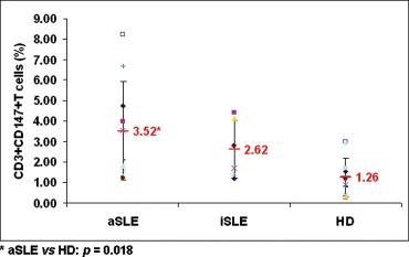
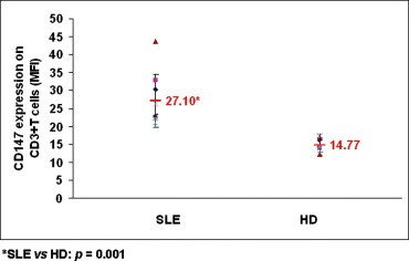
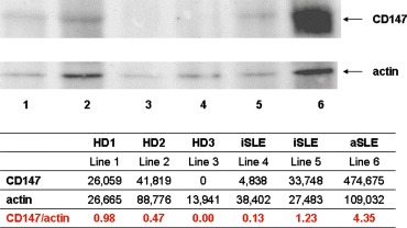
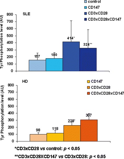
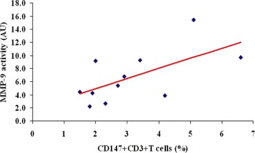
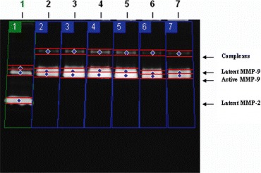
The cellular and molecular mechanisms involved in many abnormalities described in Systemic Lupus Erythematosus (SLE) are still unclear. Some of these abnormalities referred to the hyperactivation of T lymphocytes and the enhanced secretion of MMP-9 by peripheral blood mononuclear cells (PBMCs). Therefore, in this paper we investigated the potential role of CD147 molecule in these abnormalities. Our results demonstrated that CD147 molecule is overexpressed on CD3+T lymphocytes from SLE patients when compared with CD3+T lymphocytes from healthy donors. Monoclonal anti-CD147 antibodies, MEM-M6/1 clone, were able to inhibit protein tyrosine phosphorylation only in CD3 x CD28 costimulated T lymphocytes from SLE patients. However, this monoclonal antibody was unable to inhibit the enhanced activity of MMP-9 secreted by SLE PBMCs.


Figures

1
1
FACS analysis of CD3+CD147+T lymphocytes. PBMCs freshly isolated from one subject were double stained using FITC conjugated monoclonal anti-CD147 antibodies (FL1-H) and PE conjugated monoclonal anti-CD3 antibodies (FL2-H). CD3+T lymphocytes were gated (A, gate R1). The percentage of CD3+CD147+T lymphocytes from R1 is identified in upper right quadrant (B).
2
2
The percentage of CD147 positive cells in CD3+T lymphocytes from SLE patients and healthy donors. Points represent the percentages of CD3+CD147+T lymphocytes obtained for each active (aSLE) and inactive (iSLE) SLE patient and healthy donor (HD) as well as the mean values ± SD calculated for each group of subjects.
3
3
FACS analysis for the density of CD147 molecules on CD3+T lymphocytes from SLE patients and healthy donors. Median of fluorescence intensities (MFI) that corresponds to the density of CD147 molecules on CD3+T lymphocytes is presented as points for each studied subject. The mean values ± SD of MFI for each group of subjects are included in histogram.
4
4
Immunoblotting analysis of CD147 expression on PBMCs. PBMCs isolated from three healthy donors and three SLE patients (two in inactive and one in active stage of disease) were analyzed by immunoblotting using specific anti-CD147 and anti-actin antibodies. Immunoblotting was analyzed by densitometry and the expression level of CD147 on PBMCs was expressed as the ratio between CD147 and actin band intensities.
5
5
Tyrosine phosphorylation pattern in unstimulated and stimulated T lymphocytes. PBMCs isolated from one SLE patient (A) and one healthy donor (B) were stimulated with different types of monoclonal antibodies, as follows: (1) unstimulated, (2) stimulated by anti-CD147 antibodies, (3) stimulated by anti-CD3 and anti-CD28 antibodies, (4) costimulated by antibodies directed to CD3, CD28 and CD147 receptors. After 5 min of stimulation, the cells were lysed and analyzed by immunoblotting with monoclonal anti-phosphotyrosine antibodies and ECL system. The standards of known molecular weight (MW) were included in each experiment.
6
6
Tyrosine phosphorylation level in unstimulated and stimulated T lymphocytes. PBMCs isolated from SLE patients and healthy donors were stimulated as in Figure 5 and analyzed by densitometry for total tyrosine phosphorylation level. For each type of stimulation and for SLE (A) and healthy donors (B) group the mean values ± SD was calculated and indicated on histograms.
7
7
Correlation between the percentage of CD3+CD147+T lymphocytes and the activity of secreted MMP-9. PBMCs freshly isolated from peripheral blood of SLE patients were analyzed by FACS in order to determine the percentage of CD3+CD147+T lymphocytes. In addition, PBMCs from the same patients were cultured for 24 hrs and the culture supernatants were analyzed by gelatin zymography. Zymograms were scanned and the intensity of bands was determined. Subsequently, these two parameters were correlated by Spearman's analysis.
8
8
Modulation of MMPs activity in PBMCs cultured in absence or presence of monoclonal anti-CD147 antibodies. PBMCs of one SLE patient were cultured in absence (Line 2) or presence of anti-CD147 antibodies immobilized by rabbit anti-mouse IgG antibodies (Lines 4, 5) or soluble (lines 6, 7). Two controls were included in all experiments: positive control (fetal calf serum as source of latent and active forms of MMP-9 and latent MMP-2) (Line 1) and negative control (supernatant of PBMCs cultured only in the presence of rabbit antimouse IgG antibodies) (Line 3). After 24 hours, supernatants were collected and analyzed by gelatin zymography.

Similar articles

Cited by

References

    1. Kammer GM, Perl A, Richardson BC, Tsokos GC. Abnormal T cell signal transduction in systemic lupus erythematosus. Arthritis Rheum. 2002;46:1139–54. - PubMed
    1. Blasini AM, Brundula V, Paris M, Rivas L, Salazar S, Stekman IL, Rodriguez MA. Protein tyrosine kinase activity in T lymphocytes from patients with systemic lupus erythematosus. J Autoimmun. 1998;11:387–93. - PubMed
    1. Matache C, Stefanescu M, Onu A, Tanaseanu S, Matei I, Frade R, Szegli G. p56lck activity and expression in peripheral blood lymphocytes from patients with systemic lupus erythematosus. Autoimmunity. 1999;29:111–20. - PubMed
    1. Nambiar MP, Enyedy EJ, Fisher CU, Krishnan S, Warke VG, Gilliland WR, Oglesby RJ, Tsokos GC. Abnormal expression of various molecular forms and distribution of T cell receptor zeta chain in patients with systemic lupus erythematosus. Arthritis Rheum. 2002;46:163–74. - PubMed
    1. Nambiar MP, Krishnan S, Warke VG, Tsokos GC. TCR zeta-chain abnormalities in human systemic lupus erythematosus. Methods Mol Med. 2004;102:49–72. - PubMed

Publication types

MeSH terms