. 2007 May 8:5:4.
doi: 10.1186/1477-3155-5-4.

Potential therapeutic application of gold nanoparticles in B-chronic lymphocytic leukemia (BCLL): enhancing apoptosis

Affiliations

Potential therapeutic application of gold nanoparticles in B-chronic lymphocytic leukemia (BCLL): enhancing apoptosis

Priyabrata Mukherjee et al. J Nanobiotechnology. .

Erratum in

  • J Nanobiotechnology. 2013;11:23

Abstract

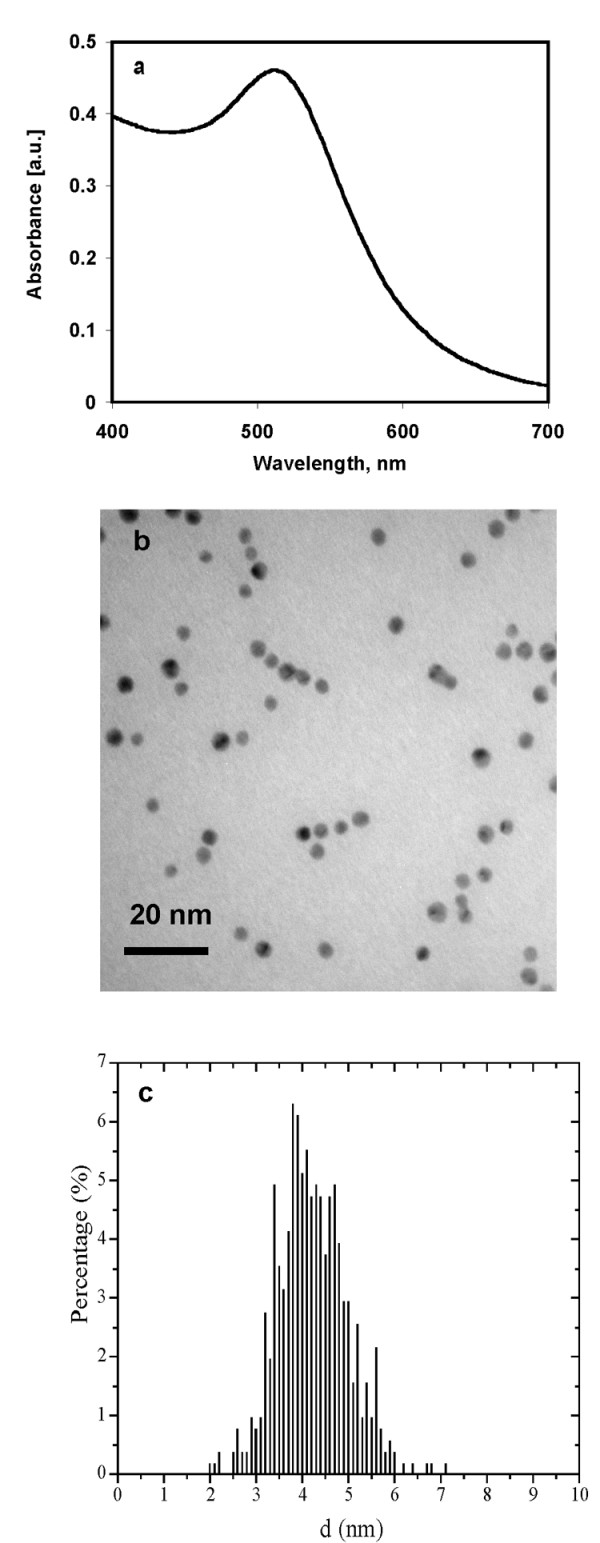
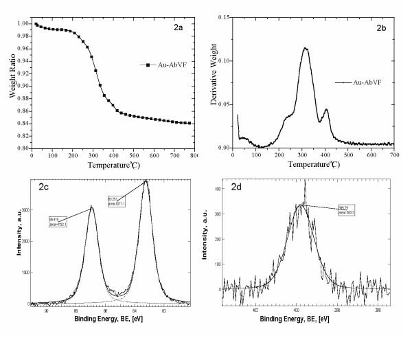
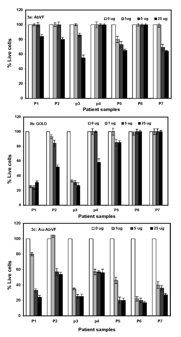
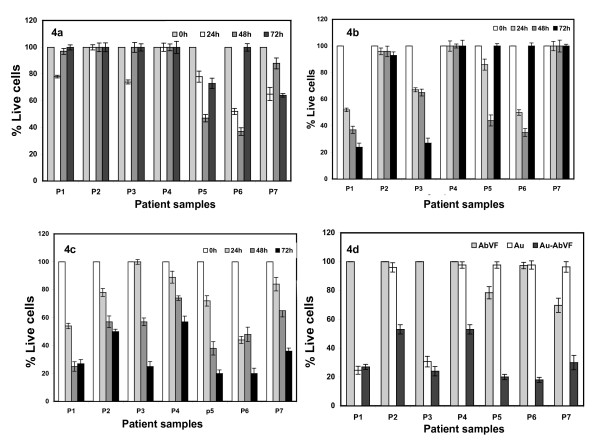
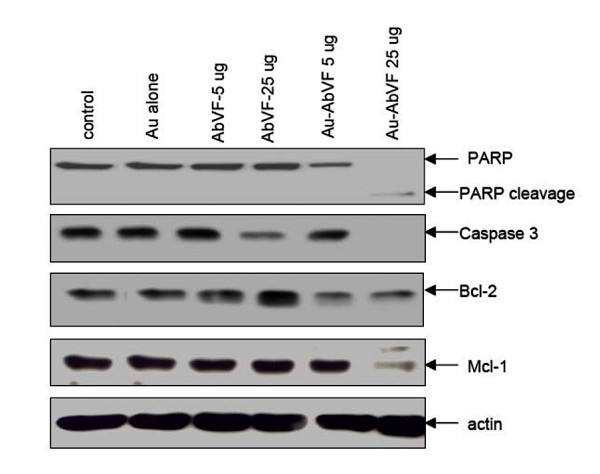
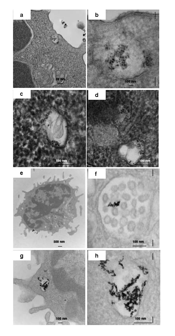
B-Chronic Lymphocytic Leukemia (CLL) is an incurable disease predominantly characterized by apoptosis resistance. We have previously described a VEGF signaling pathway that generates apoptosis resistance in CLL B cells. We found induction of significantly more apoptosis in CLL B cells by co-culture with an anti-VEGF antibody. To increase the efficacy of these agents in CLL therapy we have focused on the use of gold nanoparticles (GNP). Gold nanoparticles were chosen based on their biocompatibility, very high surface area, ease of characterization and surface functionalization. We attached VEGF antibody (AbVF) to the gold nanoparticles and determined their ability to kill CLL B cells. Gold nanoparticles and their nanoconjugates were characterized using UV-Visible spectroscopy (UV-Vis), transmission electron microscopy (TEM), thermogravimetric analysis (TGA) and X-ray photoelectron spectroscopy (XPS). All the patient samples studied (N = 7) responded to the gold-AbVF treatment with a dose dependent apoptosis of CLL B cells. The induction of apoptosis with gold-AbVF was significantly higher than the CLL cells exposed to only AbVF or GNP. The gold-AbVF treated cells showed significant down regulation of anti-apoptotic proteins and exhibited PARP cleavage. Gold-AbVF treated and GNP treated cells showed internalization of the nanoparticles in early and late endosomes and in multivesicular bodies. Non-coated gold nanoparticles alone were able to induce some levels of apoptosis in CLL B cells. This paper opens up new opportunities in the treatment of CLL-B using gold nanoparticles and integrates nanoscience with therapy in CLL. In future, potential opportunities exist to harness the optoelectronic properties of gold nanoparticles in the treatment of CLL.


Figures

Figure 1
Figure 1
UV-Visible spectroscopy and transmission electron micrograph of gold nanoparticles. a) UV-visible spectrum of a gold nanoparticles solution and b) Transmission electron microscopy picture of gold nanoparticles. TEM was done after drop coating the gold nanoparticles on carbon coated copper grid, c) histogram showing the size distribution of gold nanoparticles.
Figure 2
Figure 2
Thermogravimetric analysis (TGA) of Au-AbVF conjugates. For TGA, 150 ml gold nanoparticles were incubated with 600 μg of AbVF. After 1 h, the nanoconjugates were centrifuged at 25000 rpm for 1 h, freeze-dried overnight and analyzed using TGA; 2a) describes weight loss over temperature and 2b) derivative weight loss over temperature for the nanoconjugates. TGA analysis was done on purified and lyophilized nanoconjugates, 2c) X-ray photoelectron spectra of Au-AbVF conjugates. Core level BE of Au and 2d) core level BE of N.
Figure 3
Figure 3
Dose dependent effect of AbVF, Au and Au-AbVF in the induction of apoptosis in CLL-B cells. Cells were treated with 1, 5 and 25 ug of AbVF/Au/Au-AbVF for 72 h followed by measurement of apoptosis using annexin PI. The % apoptosis was calculated after normalizing the apoptosis in the control cells (no treatment group) to zero; 3a) effect of AbVF to induce apoptosis in CLL-B cells; 3b) effect of Au nanoparticles only to induce apoptosis in CLL-B and figure 3c) effect of Au-AbVF to induce apoptosis in CLL B cells. In the case of AbVF treated group, the amount of AbVF used is exactly the same in the corresponding doses of Au-AbVF. Similarly, in case of gold treated group, the amount of gold nanoparticles used is exactly the same as in the corresponding doses of Au-AbVF.
Figure 4
Figure 4
Effect of time on the induction of apoptosis of CLL-B cells by AbVF, Au and Au-AbVF. Cells were treated with 5 ug/ml of AbVF/Au/Au-AbVF for 72 h. Apoptosis measurement was then done using annexin/PI and apoptosis was calculated as described above; 4a) effect of AbVF alone; 4b) effect of Au alone; 4c) effect of Au-AbVF alone and 3d) comparison of the efficacy of AbVF, gold and gold-AbVF, 4d) comparison of effect of AbVF, Au and Au-AbVF on the apoptosis of CLL-B cells. To compare the activity of AbVF either conjugated or free the amount of AbVF used as control is exactly the same amount of AbVF present in the nanoconjugate. Similarly, to compare the activity of Au and Au-AbVF the amount of Au used as control is exactly the same amount of Au present in the nanoconjugate. The quantitations of Au in the nanoparticles solution and in the purified nanoconjugates were determined using inductively couple plasma analysis (ICP).
Figure 5
Figure 5
Immunoblot analysis of CLL B cells exposed to gold particles (Au) alone, AbVF alone at 5 and 25 μg/ml and AU-AbVF at 5 and 25 μg/ml. Note the prominent PARP cleavage, decrease in caspase 3, Mcl-1 and moderate change in Bcl-2 for CLL B cells treated with Au-AbVF at 25 μg/ml. These changes were noted but less evident for Au-AbVF at the 5 μg/ml dose.
Figure 6
Figure 6
Internalization of Au-AbVF and Au alone by primary CLL-B cells after 1 h incubation. 6a) nanoparticles were seen at the cell periphery (within uncoated tubules and vacuoles); 6b) higher magnification image of 6a; 6c and 6d) showing the internalized particles in different endocytic compartments. Figure 6e to 6h show the internalization of Au nanoparticles alone by primary CLL-B cells after 1 h incubation. 6e) Nanoparticles were seen at the cell periphery (within uncoated tubules and vacuoles); 6f) higher magnification image of 6e; 6f) showing the internalized particles in multivesicular bodies; and 6g) higher magnification image of 6f.

References

    1. Folkman J. Angiogenesis and apoptosis. Semin Cancer Biol. 2003;13:159–167. doi: 10.1016/S1044-579X(02)00133-5. - DOI - PubMed
    1. Mesters RM. Angiogenesis in hematologic malignancies. Ann Hematol. 2002;81:S72–S74. - PubMed
    1. Lee YK, Shanafelt TD, Bone ND, Strege AK, Jelinek DF, Kay NE. VEGF receptors on chronic lymphocytic leukemia (CLL) B cells interact with STAT 1 and 3: implication for apoptosis resistance. Leukemia. 2005;19:513–23. - PubMed
    1. List AF. Vascular endothelial growth factor signaling pathway as an emerging target in hematologic malignancies. Oncologist. 2001;6:24–31. doi: 10.1634/theoncologist.6-suppl_5-24. - DOI - PubMed
    1. Shanafelt TD, Kay NE. The clinical and biologic importance of neovascularization and angiogenic signaling pathways in chronic lymphocytic leukemia. Semin Oncol. 2006;33:174–85. doi: 10.1053/j.seminoncol.2006.01.008. - DOI - PubMed

LinkOut - more resources