. 2007 Jun 15;306(2):636-45.
doi: 10.1016/j.ydbio.2007.03.525. Epub 2007 Apr 5.

Impaired male fertility and atrophy of seminiferous tubules caused by haploinsufficiency for Foxa3

Affiliations

Impaired male fertility and atrophy of seminiferous tubules caused by haploinsufficiency for Foxa3

Rüdiger Behr et al. Dev Biol. .

Abstract

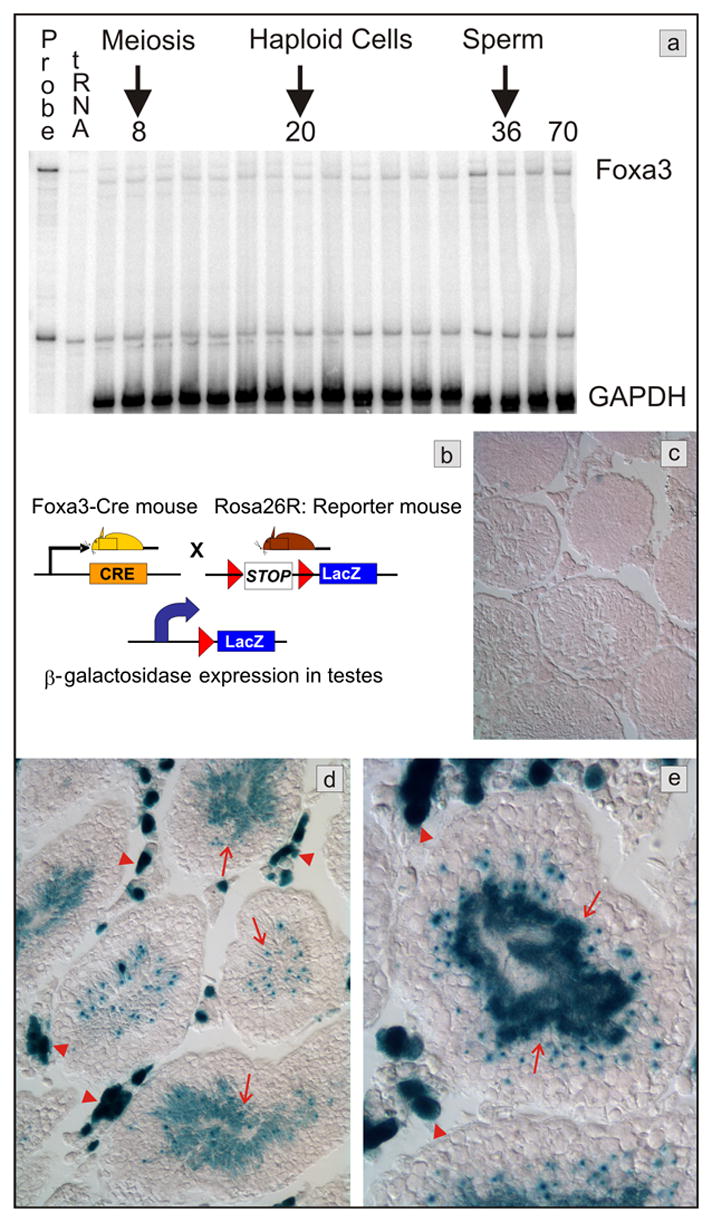
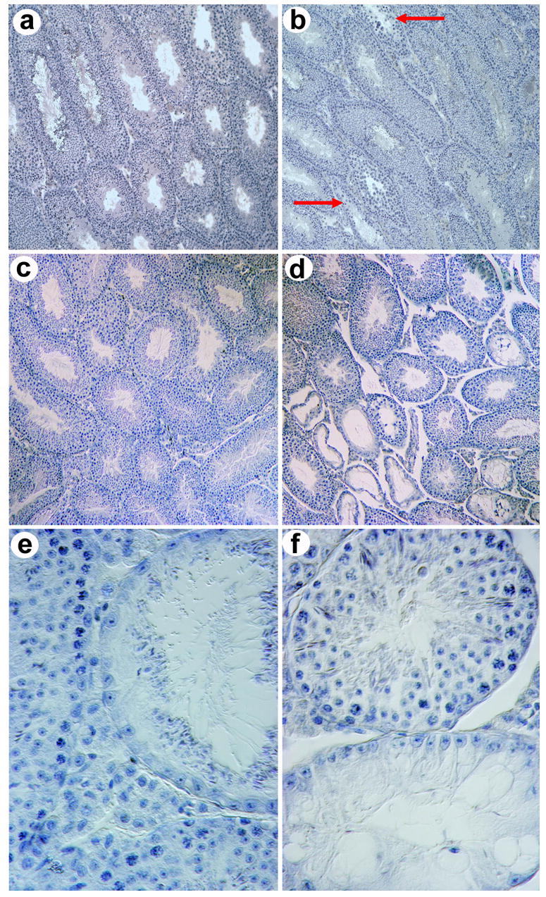
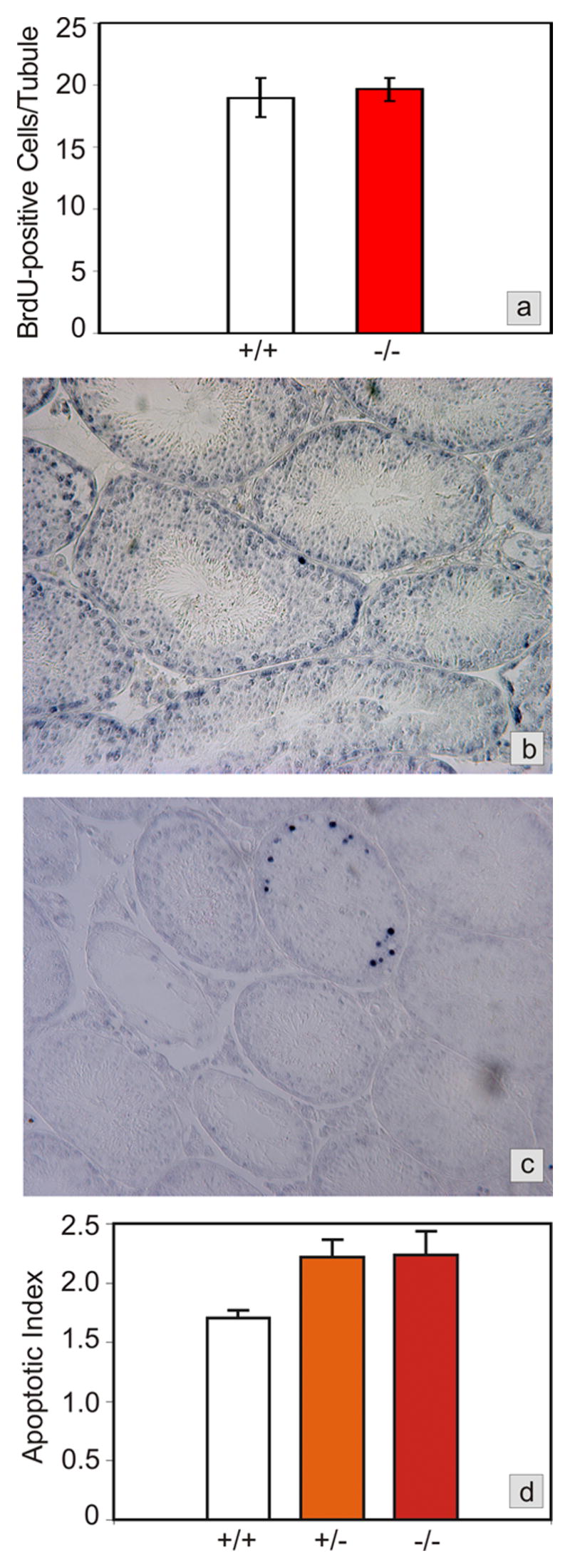
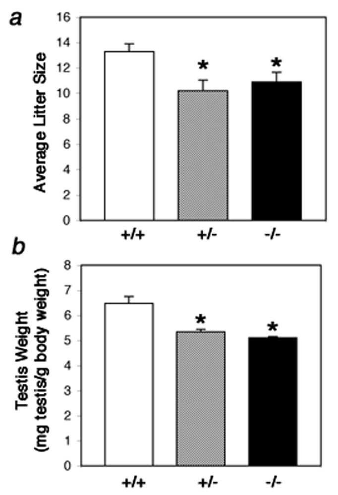
Foxa1, 2 and 3 (formerly HNF-3alpha, -beta and -gamma) constitute a sub-family of winged helix transcription factors with multiple roles in mammalian organ development. While all three Foxa mRNAs are present in endoderm derivatives including liver and pancreas, only Foxa3 is expressed in the testis. Here we demonstrate by genetic lineage tracing that Foxa3 is expressed in postmeiotic germ and interstitial Leydig cells. The germinal epithelium of Foxa3-deficient testes is characterized by a loss of germ cells secondary to an increase in germ cell apoptosis that ultimately leads to a Sertoli cell-only syndrome. Remarkably, not only the Foxa3(-/-) mice but also Foxa3(+/-) mice exhibited loss of germ cells. This cellular phenotype caused significantly reduced fertility and testis weight of both Foxa3(-/-) and Foxa3(+/-) mice. Using microarray analysis, we found a dramatic downregulation of the zinc finger protein 93 and the testicular tumor-associated paraneoplastic Ma antigen (PNMA) and increased expression of a number of genes including zinc finger protein 94 and several kallikrein 1-related peptidases which could account for at least part of the observed phenotype. In summary, we have identified Foxa3 as a transcriptional regulator with a dominant phenotype in germ cell maintenance and suggest FOXA3 as a potential candidate gene for subfertility in man.


Conflict of interest statement

Conflict of interest: The authors have declared that no conflict of interest exists.

Figures

Fig. 1
Fig. 1
Testicular expression of Foxa3. a) Messenger RNA expression analysis by RNase protection assay. Foxa3 mRNA is detectable in the mouse testis throughout postnatal development. The mRNA abundance is increased approximately 2-fold in the adult testis when compared to the 6 days old testis (normalized to GAPDH mRNA). RNA was prepared from testis of postnatal day 6, 8, 10, 12, 14, 16, 18, 20, 22, 24, 26, 28, 30, 32, 36, 40, and 70, respectively. b) Scheme of the genetic lineage tracing employed to track Foxa3 expression in the testis. Beta-galactosidase expression occurs only in those cells that express Foxa3 or are the descendants of Foxa3-expressing cells. c) The control mouse (Rosa26R, no Cre), shows no β-galactosidase expression in the testes. d, e) Mice carrying both the β-galactosidase reporter and the Foxa3-Cre transgene (Rosa26R; Foxa3Cre mice) display robust staining in Leydig cells (red arrowheads) and in spermatids (arrows).
Fig. 2
Fig. 2
Testicular degeneration in Foxa3-deficient mouse testes. a) Histological section of a wild type mouse testis at the age of three months. All seminiferous tubules contain somatic Sertoli cells as well as germ cells and exhibit ongoing germ cell production. b) Mutant testis at the age of three months. Sporadic tubulus with early signs of degeneration, i.e. thinning of the germinal epithelium, can be seen (arrow), c) Histological section of a wild type mouse testis at the age of eight months showing normal spermatogenesis in all tubules. d,e,f) Histological sections of Foxa3-deficient mouse testes at eight months of age. The testis contains both tubules with normal appearance and ongoing spermatogenesis as well as severely degenerated tubules. The characteristic feature of the defective tubules is a selective loss of germ cells. The Sertoli cells remained within the tubule resulting in a focal Sertoli-cell-only syndrome. e) An almost completely atrophied seminiferous tubule. The only germ cells present are elongated spermatids. All spermatogonia, spermatocytes, and round spermatids are missing. The elongated spermatids are arrayed on the adluminal surface of the Sertoli cells as they usually are shortly before sperm release from the germinal epithelium (spermiation). f) A Sertoli cell only tubule (bottom) adjacent to a tubule with complete spermatogenesis (top). The Sertoli cell nuclei characterized by their triangular shape and their prominent nucleoli have aligned with the basal lamina indicating the absence of any germ cells. Heterozygous testes show histological pictures similar to the homozygous mutant testes. Magnification of a), b), c), and d) 100x; e) and f) 600x.
Fig. 3
Fig. 3
Testicular proliferation and apoptosis in Foxa3-deficient mice. a) Proliferation rates, as determined by BrdU labeling of cells in S-phase during spermatogenic stages VII and VIII, are not different between wild type and Foxa3-deficient mice (p>0.35). b) Apoptosis in a wild type mouse testis as revealed by the TUNEL-technique. Only a minor proportion of all tubules contains one or more TUNEL-positive cells. All other tubules are devoid of TUNEL-positive cells. As judged from the appearance and the location of the cells in the seminiferous tubule the apoptotic cells are almost exclusively spermatogonia and spermatocytes. c) Foxa3-deficient mice loose their germ cell population by apoptosis. The tubule undergoing degeneration exhibits a high number of apoptotic germ cells, again mainly spermatogonia and spermatocytes. Note the already degenerated tubules which are devoid of TUNEL-positive cells. d) Apoptotic index expressed as TUNEL-positive cells per tubule containing TUNEL-positive cells. The mutant as well as the heterozygous testes show a significant increase in the number of apoptotic cells (p<0.05).
Fig. 4
Fig. 4
Reduced male fertility and testis weight in Foxa3-deficient mice. a) The testicular defect in Foxa3-deficient mice results in reduced fertility. Male age-matched mice of each genotype were housed together with a female CD1 wt mouse. The average litter size in the mutant (p<0.05) and in the heterozygous group (p<0.02) was significantly lower than in the wt group. b) The Foxa3-mutant (p=0.003) as well as the heterozygous (p=0.007) mice show a significantly reduced testis weight compared to the wild type controls. Data are presented as paired testis weight [mg] per body weight [g].
Fig. 5
Fig. 5
Foxa3 binding sites in the putative promotors of genes showing reduced steady state mRNA levels in Foxa3-mutant testes. Binding sites are shown in color. Numbers on the right give the position relative to the transcriptional start site. For 0710005I19Rik, there are 2 overlapping sites. ATATATATGTTT (starts in green, ends in light blue) and ATATGTTTATGT (starts in light blue, ends in dark blue). The part in light blue is the overlap (ATATGTTT). For all others the full predicted sites (12 bp) are in color. TGTTT is the core motif for Foxa binding.
Fig. 6
Fig. 6
Degenerating seminiferous tubules in Foxa3-deficient testes re-express Anti-Muellerian hormone (AMH) as revealed by immunohistochemistry (a–d). a) A seminiferous tubule in the early phase of degeneration. The thickness of the germinal epithelium appears to be normal, although vacuolization of the epithelium has already occurred. Germ cells present in this tubule are mostly round spermatids as judged from nuclear morphology. Earlier germ cell stages (spermatogonia and spermatocytes) are rather rare. Germ cells are devoid of any pink stain (yellow arrows) but the surrounding cytoplasm of the Sertoli cells is clearly stained (red arrows). Tubules with normal appearance do not exhibit any staining (lower left part). b) A degenerating tubule in a more advanced stage than that shown in a). One generation of spermatocytes (presumably maturing to spermiation) is still present in this tubule. Sertoli cell nuclei have aligned along the tubular wall and the cytoplasm is clearly stained by the AMH antibody (red arrow) c) A tubule during degeneration (red asterisk) still containing some germ cells (yellow arrows) and a tubule at the final stage of degeneration (yellow asterisk), which exhibits a Sertoli cell only-phenotype characterized by the complete loss of germ cells. At this stage the Sertoli cells have flattened and extend little beyond their nuclei. Again, AMH immunoreactivity is widespread. Red arrows point out AMH positive Sertoli cells. d) Negative control omitting the AMH antibody from the diluent. Also, the adult wild type control mice exhibited no staining while Sertoli cells in young testes up to p10 showed strong cytoplasmic Sertoli cell staining (data not shown). e) Expression of the androgen receptor (AR; brown stain) in a wt testis as revealed by immunohistochemistry. Sertoli cells (long red arrows), peritubular myoid cells (short red arrows) and Leydig cells (red asterisk) are AR positive. f) AR expression in an 8 months old mutant testis showing staining in the same cell types as the age-matched wildtype control. Remarkably, also Sertoli cells in almost completely degenerated tubules still express the AR.

References

    1. Andersson AM, Jorgensen N, Frydelund-Larsen L, Rajpert-De Meyts E, Skakkebaek NE. Impaired Leydig cell function in infertile men: a study of 357 idiopathic infertile men and 318 proven fertile controls. J Clin Endocrinol Metab. 2004;89:3161–7. - PubMed
    1. Behr R, Weinbauer GF. Germ cell-specific cyclic adenosine 3′,5′-monophosphate response element modulator expression in rodent and primate testis is maintained despite gonadotropin deficiency. Endocrinology. 1999;140:2746–54. - PubMed
    1. Behringer RR, Finegold MJ, Cate RL. Mullerian-inhibiting substance function during mammalian sexual development. Cell. 1994;79:415–25. - PubMed
    1. Blaber M, Isackson PJ, Bradshaw RA. A complete cDNA sequence for the major epidermal growth factor binding protein in the male mouse submandibular gland. Biochemistry. 1987;26:6742–9. - PubMed
    1. Clements JA, Willemsen NM, Myers SA, Dong Y. The tissue kallikrein family of serine proteases: functional roles in human disease and potential as clinical biomarkers. Crit Rev Clin Lab Sci. 2004;41:265–312. - PubMed

Publication types