. 2007 May 15;104(20):8293-8.
doi: 10.1073/pnas.0700402104. Epub 2007 May 8.

Structural basis for functional mimicry of long-variable-arm tRNA by transfer-messenger RNA

Affiliations

Structural basis for functional mimicry of long-variable-arm tRNA by transfer-messenger RNA

Yoshitaka Bessho et al. Proc Natl Acad Sci U S A. .

Abstract

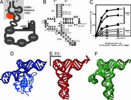
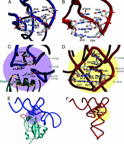
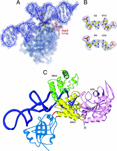
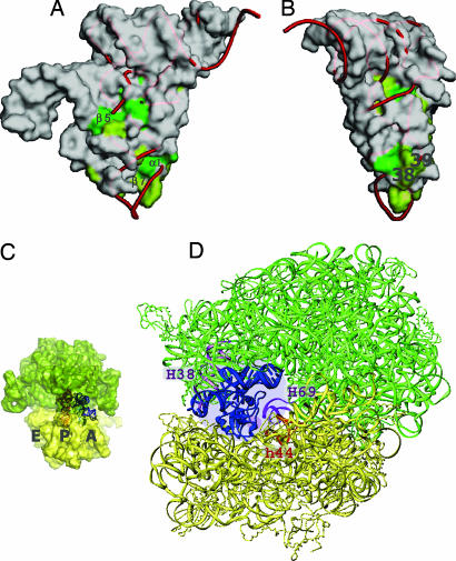
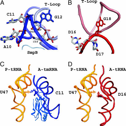
tmRNA and small protein B (SmpB) are essential trans-translation system components. In the present study, we determined the crystal structure of SmpB in complex with the entire tRNA domain of the tmRNA from Thermus thermophilus. Overall, the ribonucleoprotein complex (tRNP) mimics a long-variable-arm tRNA (class II tRNA) in the canonical L-shaped tertiary structure. The tmRNA terminus corresponds to the acceptor and T arms, or the upper part, of tRNA. On the other hand, the SmpB protein simulates the lower part, the anticodon and D stems, of tRNA. Intriguingly, several amino acid residues collaborate with tmRNA bases to reproduce the canonical tRNA core layers. The linker helix of tmRNA had been considered to correspond to the anticodon stem, but the complex structure unambiguously shows that it corresponds to the tRNA variable arm. The tmRNA linker helix, as well as the long variable arm of class II tRNA, may occupy the gap between the large and small ribosomal subunits. This suggested how the tRNA domain is connected to the mRNA domain entering the mRNA channel. A loop of SmpB in the tRNP is likely to participate in the interaction with alanyl-tRNA synthetase, which may be the mechanism for the promotion of tmRNA alanylation by the SmpB protein. Therefore, the tRNP may simulate a tRNA, both structurally and functionally, with respect to aminoacylation and ribosome entry.


Conflict of interest statement

The authors declare no conflict of interest.

Figures

Fig. 1.
Fig. 1.
Structural mimicry of a long-variable-arm tRNA by tmRNA with SmpB. (A) Schematic diagram showing the secondary structure of tmRNA. The tRNA domain is highlighted in a shaded square. The SmpB-binding site is colored orange. PK means pseudoknot structure of RNA. The asterisk shows the stop codon of the short mRNA. (B) The secondary structure of the tRNA domain of T. thermophilus tmRNA. The substituted base-pairs in the P2a and P10 stems, and the connecting UUCG tetra-loop are shaded. The actual nucleotide numbers for the 3′-terminus of the tmRNA are in parentheses. The squares indicate the nucleotides involved in the interaction between the T and D loops. Triangles show nucleotides that interact with SmpB (SI Fig. 6B and SI Table 2). (C) Alanylation of truncated tmRNAs by alanyl-tRNA synthetase, in the presence (closed symbols) and the absence (open symbols) of SmpB. (D) Overall structure of the entire tRNA domain of tmRNA complexed with SmpB, determined in this study. (E and F) Yeast tRNAPhe (33) and T. thermophilus tRNASer (10) represent short (class I) and long (class II) variable-arm tRNAs, respectively. The disordered acceptor stem, anticodon and variable-loop structures of tRNASer were completed by using yeast tRNAPhe and a typical 6nt loop (34).
Fig. 2.
Fig. 2.
Detailed structures of tmRNA and SmpB, compared with yeast tRNAPhe. The tmRNA and SmpB are colored blue and blue-green, respectively (A, C, and E), and the tRNAPhe is red (B, D, and F). (A and B) The T loop and D loop connection for tmRNA and yeast tRNAPhe are viewed from an appropriate angle to show the base-stacking. The acceptor and T stems are in the upper areas of both figures. (C and D) The base stacking of the central core is shown for tmRNA and yeast tRNAPhe, respectively. The amino acid residues of SmpB, which participate in the base stacking, are labeled in light-yellow in C. (E and F) The regions of the central cores are shown on the overall structure of tmRNA (purple) and tRNAPhe (yellow). Both figures (E and F) are horizontally rotated by 30° from C and D, respectively. The central loop of SmpB is colored red (E). The structure of yeast tRNAPhe is the most suitable to compare the detailed structure with tmRNA, among all of the known tRNA structures to date. The long-variable-arm (class II) tRNAs, despite their lower resolution, have almost the same structure as that of yeast tRNAPhe in both the T loop to D loop connection and the central core region, besides a base (position 46) in the long-variable arm (10, 35, 36).
Fig. 3.
Fig. 3.
Alanyl-activation model of tmRNA caused by SmpB. (A) Stick model of tmRNA-TDc in complex with SmpB, shown on the electron density map (contour level 1.0 σ). The nucleotides and amino acid residues of the central core region are depicted by the yellowish elemental models. The residues (from Tyr-63 to Asn-70) in the central loop of SmpB are colored red near the lower acceptor stem of tmRNA. (B) The G3-U70 (343) wobble base pair, which is the most important aminoacyl-discriminator by AlaRS, is compared with the G4-C69 (342) Watson–Crick base pair. (C) Docking model of the tmRNA-TDc with SmpB and the N-domain of A. aeolicus AlaRS (14). The tmRNA-TDc and SmpB are colored blue and blue-green, respectively. The central loop of SmpB is red. For AlaRS, three domains (ASD, active site domain; RRD1, RNA recognition domain 1; RRD2, RNA recognition domain 2) are colored pink, yellow, and green, respectively. This model is an imitation of the original superimposed method for AlaRS and tRNA (14); that is, it represents individual superpositions of AlaRS (PDB ID code 1RIQ) and tmRNA-TDc, on the structure of the complex of AspRS and tRNAAsp (PDB ID code 1C0A) (37).
Fig. 4.
Fig. 4.
Functional mimicry of a canonical tRNA by tmRNA and SmpB. (A and B) Molecular surface of SmpB, represented as with tmRNA. The conserved residues on the surface of SmpB are colored green (complete) or light green (partial). The tRNAPhe was superimposed on the tRNA domain of the tmRNA by using the nucleotides of the acceptor stems and the T arms, and the central cores with the residues of SmpB corresponding to the nucleotides (Fig. 2C). Positions 38 and 39 of the tRNA are labeled in B. (C and D) tmRNA with SmpB on the ribosome. The 50S and 30S subunits are colored green and yellow, respectively. tmRNA and SmpB are superimposed on the A-site tRNA of the 70S ribosome (16). The yeast tRNAPhe is shown in the P-site of the ribosome in C. The corresponding region of SmpB for nucleotide positions 38 and 39, shown in A and B, is indicated by a small purple circle around H69 and h44 of the ribosome in D.
Fig. 5.
Fig. 5.
Structural function of the shortened D arm of tmRNA. (A) The A10 and C11 bases in the deficient D loop of tmRNA are shown by stick models. G12, a key base connecting the D loop with the T loop, is shown in solid blue. C59 (332) and U60 (333) of the T loop are shown to indicate that their bases stabilize the backbone of A10 and C11. (B) D16, D17, and G18 of yeast tRNAPhe are colored in the same manner as the corresponding bases of tmRNA (A). G18, which interacts with Ψ55, is colored solid red, instead of solid blue for the G12 of tmRNA. (C and D) The SmpB-bound tmRNA-TDc is superimposed with the A-site tRNA of the 70S ribosome, in the same manner as in Fig. 4 C and D. In the A site of the ribosome, the tmRNA with SmpB (C) is colored blue, and the tRNAPhe (D) is red. The P-site tRNAPhe is colored yellow in both C and D. C11 of the A-site tmRNA, which spatially corresponds to D16 of the A-site tRNAPhe, is shown as a stick model.

Similar articles

Cited by

References

    1. Keiler KC, Waller PR, Sauer RT. Science. 1996;271:990–993. - PubMed
    1. Karzai AW, Roche ED, Sauer RT. Nat Struct Biol. 2000;7:449–455. - PubMed
    1. Karzai AW, Susskind MM, Sauer RT. EMBO J. 1999;18:3793–3799. - PMC - PubMed
    1. Hanawa-Suetsugu K, Takagi M, Inokuchi H, Himeno H, Muto A. Nucleic Acids Res. 2002;30:1620–1629. - PMC - PubMed
    1. Shimizu Y, Ueda T. FEBS Lett. 2002;514:74–77. - PubMed

Publication types

Associated data

LinkOut - more resources