. 2007 May 15;104(20):8281-6.
doi: 10.1073/pnas.0702259104. Epub 2007 May 8.

Rigidity-driven growth and migration of epithelial cells on microstructured anisotropic substrates

Affiliations

Rigidity-driven growth and migration of epithelial cells on microstructured anisotropic substrates

Alexandre Saez et al. Proc Natl Acad Sci U S A. .

Abstract

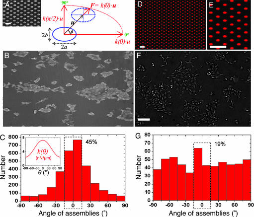
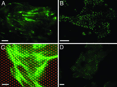
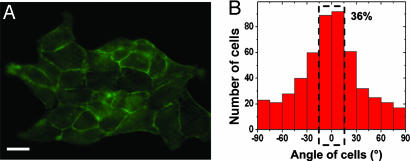
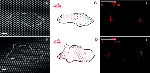
The physical properties of the cellular environment are involved in regulating the formation and maintenance of tissues. In particular, substrate rigidity appears to be a key factor dictating cell response on culture surfaces. Here we study the behavior of epithelial cells cultured on microfabricated substrates engineered to exhibit an anisotropic stiffness. The substrate consists of a dense array of micropillars of oval cross-section, so that one direction is made stiffer than the other. We demonstrate how such an anisotropic rigidity can induce directional epithelial growth and guide cell migration along the direction of greatest rigidity. Regions of high tractional stress and large cellular deformations within the sheets of cells are concentrated at the edges, in particular at the two poles of the islands along their long axis, in correlation with the orientation of actin stress fibers and focal adhesions. By inducing scattering activity of epithelial cells, we show that isolated cells also migrate along the direction of greatest stiffness. Taken together, these findings show that the mechanical interactions of cells with their microenvironment can be tuned to engineer particular tissue properties.


Conflict of interest statement

The authors declare no conflict of interest.

Figures

Fig. 1.
Fig. 1.
Epithelial cell growth on substrates with anisotropic rigidity. (A) Schematic representation of an anisotropic micropillar subjected to a force, F, where u⃗ is the displacement vector of the top of the pillar and θ is the direction of its deflection with respect to the longest semi-axis, a [i.e., the stiffest direction of the substrate (θ = 0°)]. The spring constant, k(θ), depends on the force orientation (see Eq. 1). (Inset) Scanning electron micrograph of an array of oval PDMS pillars. (Scale bar: 5 μm.) (B) MDCK cell islands grown on these substrates and visualized by optical microscopy. The horizontal direction corresponds to the polar axis, θ = 0°. (Image dimensions: length × height = 877 × 512 μm.) (C) Angular distribution of cell assemblies with respect to the stiffest direction (θ = 0°). The dashed rectangle indicates that 45% of the islands are elongated in a 30°-wide sector centered on θ = 0°. (Inset) Profile plot of the stiffness k(θ) for this experiment, computed from Eq. 1. (D) Fluorescence microscopy image of an array of oval patches of Cy3-labeled fibronectin microprinted on a glass coverslip. (Scale bar: 10 μm.) (E) Magnified view of the microprinted patches from D. (Scale bar: 10 μm.) (F) MDCK cells cultured on a microprinted coverslip. (Scale bar: 100 μm.) (G) Angular distribution of cell assemblies on patterned glass. The dashed rectangle indicates that 19% of the islands are elongated in a 30°-wide sector centered on θ = 0°.
Fig. 2.
Fig. 2.
Orientation of the actin cytoskeleton and focal adhesions on microfabricated substrates. (A and B) Immunofluorescence images of filamentous actin (A) and protein vinculin (B) in MDCK cell islands grown on anisotropic PDMS micropillars (stiffest direction, horizontal). (C) Immunofluorescence staining of filamentous actin (green) in cells grown on a glass coverslip microprinted with Cy3-fibronectin (red). (D) Immunofluorescence staining of vinculin on a fibronectin-patterned coverslip (patches oriented horizontally). (Scale bars: 10 μm.)
Fig. 3.
Fig. 3.
Orientation of individual cells within the islands. (A) Immunofluorescence staining of cortical actin showing cell–cell junctions in a MDCK cell island. (Scale bar: 20 μm.) (B) Histogram of the angular orientation of individual cells within the islands. The dashed rectangle indicates that 36% of the islands are elongated in a 30°-wide sector centered on θ = 0°.
Fig. 4.
Fig. 4.
Traction force experiments. (A) Fluorescence microscopy image of the tops of the micropillars coated and labeled with fibronectin-Cy3. (B) Transmission microscopy image of a cell island lying on the micropillars. The positions of the islands are outlined in white. (Scale bars: 10 μm.) (C and D) Maps of the instantaneous traction forces detected within the boundary of the islands at times corresponding to A (C) and B (D) on substrates with two different rigidities: k(θ = 0°) = 56 and 19 nN/μm, respectively. (E and F) Color maps of the average magnitude of the forces applied over a 1-h period by the cellular islands shown in A (E) and B (F), calculated from time-lapse sequences.
Fig. 5.
Fig. 5.
Effects of anisotropic rigidity on cell migration. (A) Trajectories of two cells over the course of 5-h experiments of hepatocyte growth factor-induced cell migration on pillars having a circular cross-section (solid line) and on pillars having an oval cross-section (dashed line). The starting position of the center of mass was set to (0, 0) for the two cells. (Inset) Magnified view of the path followed by the cell migrating on cylindrical pillars. (B) Histogram of migratory trajectories of individual cells on PDMS anisotropic micropillars. The histogram represents the angular distribution of the displacement vector linking the center of mass of a cell between two consecutive images (at time t and time t + 60 s) with respect to the horizontal direction. The dashed rectangle indicates that 25% of the islands are elongated in a 30°-wide sector centered on θ = 0°.

References

    1. Gilbert SF. Developmental Biology. 7th Ed. Sunderland, MA: Sinauer; 2003. pp. 3–180.
    1. Martin P. Science. 1997;276:75–81. - PubMed
    1. Bernstein LR, Liotta LA. Curr Opin Oncol. 1994;6:106–113. - PubMed
    1. Steinberg MS. Science. 1962;137:762–763. - PubMed
    1. Tarbell JM, Weinbaum S, Kamm RD. Ann Biomed Eng. 2005;33:1719–1723. - PubMed

Publication types

LinkOut - more resources