. 2007;35(11):3631-45.
doi: 10.1093/nar/gkm145. Epub 2007 May 8.

Characterization of Agrobacterium tumefaciens DNA ligases C and D

Affiliations

Characterization of Agrobacterium tumefaciens DNA ligases C and D

Hui Zhu et al. Nucleic Acids Res. 2007.

Abstract

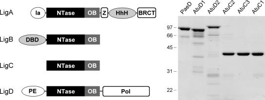
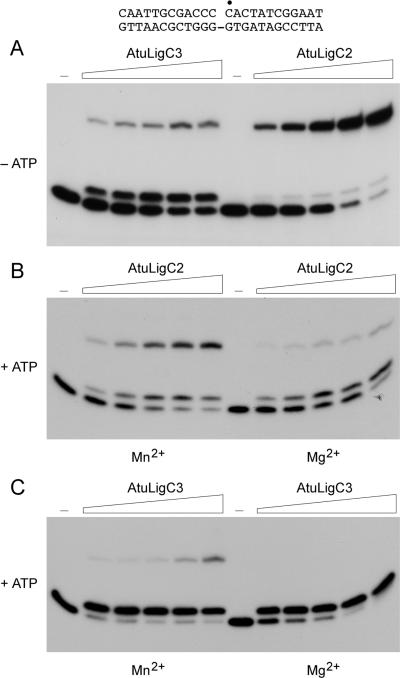
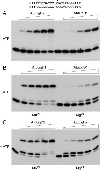
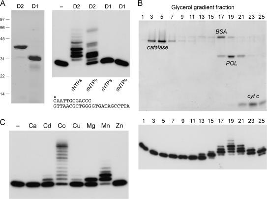
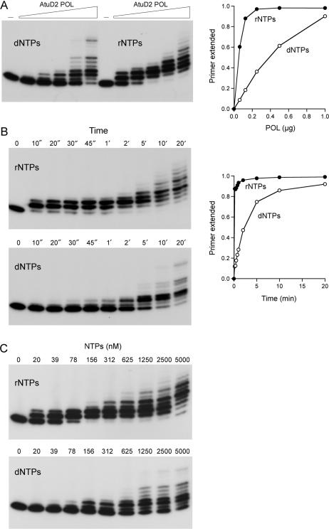
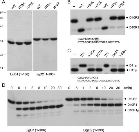
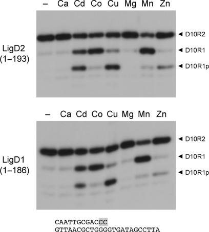
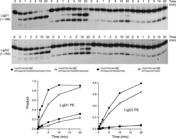
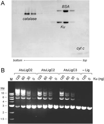
Agrobacterium tumefaciens encodes a single NAD+-dependent DNA ligase and six putative ATP-dependent ligases. Two of the ligases are homologs of LigD, a bacterial enzyme that catalyzes end-healing and end-sealing steps during nonhomologous end joining (NHEJ). Agrobacterium LigD1 and AtuLigD2 are composed of a central ligase domain fused to a C-terminal polymerase-like (POL) domain and an N-terminal 3'-phosphoesterase (PE) module. Both LigD proteins seal DNA nicks, albeit inefficiently. The LigD2 POL domain adds ribonucleotides or deoxyribonucleotides to a DNA primer-template, with rNTPs being the preferred substrates. The LigD1 POL domain has no detectable polymerase activity. The PE domains catalyze metal-dependent phosphodiesterase and phosphomonoesterase reactions at a primer-template with a 3'-terminal diribonucleotide to yield a primer-template with a monoribonucleotide 3'-OH end. The PE domains also have a 3'-phosphatase activity on an all-DNA primer-template that yields a 3'-OH DNA end. Agrobacterium ligases C2 and C3 are composed of a minimal ligase core domain, analogous to Mycobacterium LigC (another NHEJ ligase), and they display feeble nick-sealing activity. Ligation at DNA double-strand breaks in vitro by LigD2, LigC2 and LigC3 is stimulated by bacterial Ku, consistent with their proposed function in NHEJ.


Figures

Figure 1.
Figure 1.
Multiple DNA ligases of A. tumefaciens. The Atu LigA, LigB, LigC and LigD polypeptides are depicted (left panel) in cartoon form with the N-termini on the left and the C-termini on the right. The core ligase catalytic domains, composed of nucleotidyltransferase (NTase) and OB modules, are shown as rectangles. Flanking domains of known structure or imputed function (variously drawn as ellipses or capsules) are discussed in the text. Aliquots (5 µg) of the nickel-agarose preparations of PaeLigD, AtuLigD1, AtuLigD2, AtuLigC2, AtuLigC3 and AtuLigC1 were analyzed by SDS-PAGE. The Coomassie blue-stained gel is shown in the right panel. The positions and sizes (in kDa) of marker polypeptides are indicated.
Figure 2.
Figure 2.
Nick-sealing activity of AtuLigC2 and AtuLigC3. (A) Reaction mixtures (20 µl) containing either 50 mM Tris-HCl (pH 7.5) (AtuLigC2) or 50 mM Tris-acetate (pH 6.0) (AtuLigC3), 5 mM DTT, 5 mM MnCl2, 1 pmol 32P-labeled nicked 24-mer DNA substrate (depicted at the top with the labeled 5′ nucleotide at the nick indicated by filled circle), and increasing amounts of AtuLigC2 (38, 75, 150, 300 and 600 ng) or AtuLigC3 (50, 100, 200, 400 and 800 ng) were incubated for 20 min at 37°C. Enzyme was omitted from a control reaction mixture (lane −). The products were resolved by PAGE and visualized by autoradiography. (B) Reaction mixtures (20 µl) containing 50 mM Tris-HCl (pH 7.5), 5 mM DTT, 5 mM MnCl2 (left panel) or 5 mM MgCl2 (right panel), 250 µM ATP, 1 pmol 32P-labeled nicked 24-mer DNA substrate, and increasing amounts of AtuLigC2 (38, 75, 150, 300 and 600 ng) were incubated at 37°C for 20 min. Enzyme was omitted from a control reaction mixture (lane −). (C) Reaction mixtures (20 µl) containing 50 mM Tris-acetate (pH 6.0), 5 mM DTT, 5 mM MnCl2 (left panel) or MgCl2 (right panel), 250 µM ATP, 1 pmol 32P-labeled nicked 24-mer DNA substrate, and increasing amounts of AtuLigC3 (50, 100, 200, 400 and 800 ng) were incubated at 37°C for 20 min. Enzyme was omitted from a control reaction mixture (lane −).
Figure 3.
Figure 3.
Nick-sealing activity of AtuLigD1 and AtuLigD2. (A) Reaction mixtures (20 µl) containing either 50 mM Tris-HCl (pH 7.5) (AtuLigD1) or 50 mM Tris-acetate (pH 6.0) (AtuLigD2), 5 mM DTT, 5 mM MnCl2, 1 pmol 32P-labeled nicked 24-mer DNA substrate (shown at the top), and increasing amounts of AtuLigD1 (63, 125, 250, 500 and 1000 ng) or AtuLigD2 (31, 63, 125, 250, 500 ng) were incubated for 20 min at 37°C. (B) Reaction mixtures (20 µl) containing 50 mM Tris-HCl (pH 7.5), 5 mM DTT, 5 mM MnCl2 (left panel) or MgCl2 (right panel), 250 µM ATP, 1 pmol 32P-labeled nicked 24-mer DNA substrate, and increasing amounts of AtuLigD1 (63, 125, 250, 500, 1000 ng) were incubated at 37°C for 20 min. (C) Reaction mixtures (20 µl) containing 50 mM Tris-acetate (pH 6.0), 5 mM DTT, 5 mM MnCl2 (left panel) or MgCl2 (right panel), 250 µM ATP, 1 pmol 32P-labeled nicked 24-mer DNA substrate, and increasing amounts of AtuLigD2 (31, 63, 125, 250, 500 ng) were incubated at 37°C for 20 min. Enzyme was omitted from control reaction mixtures (lanes –).
Figure 4.
Figure 4.
Recombinant LigD2 and LigD1 POL domains. (A) Aliquots (7 µg) of the Ni-agarose preparations of the POL domains of AtuLigD2 and AtuLigD1 were analyzed by SDS-PAGE. The Coomassie blue-stained gel is shown in the left panel. Polymerase reaction mixtures (20 µl) containing 50 mM Tris-HCl (pH 7.5), 5 mM DTT, 5 mM MnCl2, 100 µM rNTPs or dNTPs as specified, 50 nM 5′ 32P-labeled 12-mer/24-mer DNA primer-template (depicted at bottom) and 1 µg of LigD2 or LigD1 POL as specified were incubated at 37°C for 20 min. The products were analyzed by denaturing PAGE. An autoradiograph of the gel is shown in the right panel. POL was omitted from the control reaction in lane −. (B) Glycerol gradient sedimentation was performed as described under Experimental Procedures. Aliquots (15 µl) of the odd-numbered fractions were analyzed by SDS-PAGE (top panel). The catalase, BSA, LigD2 POL and cytochrome c polypeptides are indicated. Polymerase activity was gauged by incubating 20 µl reaction mixtures containing 50 mM Tris-HCl (pH 7.5), 5 mM DTT, 5 mM MnCl2, 100 µM rNTPs, 1 pmol 5′ 32P-labeled 12-mer/24-mer DNA primer-template and 2 µl of the indicated gradient fractions at 37°C for 20 min. PAGE analysis of the primer extension products is shown in the bottom panel. (C) Metal specificity. Reaction mixtures (20 µl) containing 50 mM Tris-HCl (pH 7.5), 100 µM rNTPs, 50 nM 5′ 32P-labeled 12-mer/24-mer DNA primer-template, 156 nM LigD2 POL, and either no divalent cation (–) or 5 mM Ca2+, Cd2+, Co2+, Cu2+, Mg2+, Mn2+ or Zn2+ were incubated at 37°C for 20 min.
Figure 5.
Figure 5.
Preferential utilization of ribonucleotides by LigD2 POL. (A) Enzyme titration. Reaction mixtures (20 µl) containing 50 mM Tris-HCl (pH 7.5), 5 mM DTT, 5 mM MnCl2, 50 nM 5′ 32P-labeled 12-mer/24-mer DNA primer-template, 100 µM rNTPs or dNTPs as specified, and increasing amounts of LigD2 POL (62, 125, 250, 500 and 1000 ng, from left to right in each titration series, corresponding to 78, 156, 312, 625 and 1250 nM POL) were incubated at 37°C for 20 min. PAGE analysis of the primer extension products is shown in the left panel. POL was omitted from the control reaction in lane –. The fraction of input primer extended by at least one nucleotide is plotted as a function of input POL in the right panel. (B) Kinetics of primer extension. Reaction mixtures (220 µl) containing 50 mM Tris-HCl (pH 7.5), 5 mM DTT, 5 mM MnCl2, 50 nM 5′ 32P-labeled 12-mer/24-mer DNA primer-template, 100 µM rNTPs or dNTPs as specified, and 1.25 µM LigD2 POL were incubated at 37°C. Aliquots (20 µl) were withdrawn at the times specified and quenched immediately with EDTA/formamide. PAGE analysis of the primer extension products is shown in the left panels. The fraction of input primer extended by at least one nucleotide is plotted as a function of time in the right panel. (C) NTP concentration dependence. Reaction mixtures (20 µl) containing 50 mM Tris-HCl (pH 7.5), 5 mM DTT, 5 mM MnCl2, 50 nM 5′ 32P-labeled 12-mer/24-mer DNA primer-template, 1.25 µM LigD2 POL and rNTPs or dNTPs as specified were incubated at 37°C for 20 min.
Figure 6.
Figure 6.
End-healing activities of the AtuLigD1 and AtuLigD2 PE domains. (A) Aliquots (5 µg) of the Ni-agarose preparations of the wild-type (WT) and mutated versions of the N-terminal PE domains of AtuLigD1 and AtuLigD2 were analyzed by SDS-PAGE. The Coomassie blue-stained gel is shown. The positions and sizes (kDa) of marker polypeptides are indicated on the left. (B) Reaction mixtures (10 µl) containing 50 mM Tris-acetate (pH 6.0), 5 mM DTT, 0.5 mM MnCl2, 50 nM pmol 32P-labeled D10R2 primer-template (shown at bottom, with ribonucleotides highlighted in shaded boxes), and 4 µM WT or mutant PE domain as specified were incubated at 37°C for 20 min. The products were resolved by PAGE and visualized by autoradiography. The labeled species corresponding to the D10R2 substrate and the D10R1 end-product are indicated by arrowheads on the right. (C) Reaction mixtures (10 µl) containing 50 mM Tris-acetate (pH 6.0), 5 mM DTT, 0.5 mM MnCl2, 50 nM 32P-labeled D11p primer-template (shown at bottom) and 4 µM WT or mutant PE domain as specified were incubated at 37°C for 20 min. The products were analyzed by PAGE and visualized by autoradiography. The labeled species corresponding to the D11p substrate and the D11OH product are indicated by arrowheads on the right. (D) Reaction mixtures (90 µl) containing 50 mM Tris-acetate (pH 6.0), 5 mM DTT, 0.5 mM MnCl2, 100 nM 32P-labeled D10R2 primer-template and 4 µM AtuLigD1 or AtuLigD2 PE as specified were incubated at 37°C. Aliquots (10 µl) were withdrawn at the times specified above the lanes and quenched immediately with EDTA/formamide. The products were resolved by PAGE and visualized by autoradiography. The labeled species corresponding to the D10R2 substrate, the D10R1p intermediate and the D10R1 end-product are indicated by arrowheads on the right.
Figure 7.
Figure 7.
Divalent cation specificity of the 3′-ribonuclease activity of AtuLigD PE domains. Reaction mixtures (10 µl) containing 50 mM Tris-acetate (pH 6.0), 100 nM 32P-labeled D10R2 primer-template, 0.5 mM divalent cation as specified, and either 1 µM AtuLigD1 PE (bottom panel) or 2 µM AtuLigD2 PE (top panel) were incubated at 37°C for 20 min.
Figure 8.
Figure 8.
Effect of the template strand 5′ tail on AtuLigD PE activity. Reaction mixtures (80 µl) containing 50 mM Tris-acetate (pH 6.0), 5 mM DTT, 0.5 mM MnCl2, 50 nM 32P-labeled D10R2 primer-templates with variable 5′ tails and 4 µM AtuPE1 or AtuPE2 were incubated at 37°C. Aliquots (10 µl) were withdrawn at the times specified and quenched immediately with EDTA/formamide. The products were resolved by PAGE. The structures of the primer-templates are depicted below the autoradiograms, with ribonucleotides highlighted in shaded boxes. The kinetic profiles of the reactions were quantified by scanning the gel. Phosphodiesterase activity, expressed as the fraction of radiolabeled material in the D10R1-p plus D10R1 products, is plotted as a function of time. The substrate symbols are as depicted below the autoradiograms.
Figure 9.
Figure 9.
Ku stimulation of double-strand break repair by Atu ligases. (A) Homodimeric quaternary structure of Ku. Glycerol gradient sedimentation of PaeKu was performed as described under Experimental Procedures. Aliquots (15 µl) of odd-numbered gradient fractions were analyzed by SDS-PAGE. The catalase, BSA, Ku and cytochrome c polypeptides are indicated. (B) Plasmid end joining. Reaction mixtures (20 µl) containing 50 mM Tris-HCl (pH 7.5), 5 mM DTT, 5 mM MnCl2, 0.25 mM ATP, 0.2 µg BamHI-digested pUC19 DNA (120 fmol plasmid; 240 fmol double-strand breaks), either AtuLigD2 (250 ng), AtuLigC2 (75 ng; 1.9 pmol) or AtuLigC3 (125 ng; 3.1 pmol), and PaeKu as indicated were incubated for 2 h at 37°C. The reactions were quenched by adjusting the mixtures to 0.2% SDS and 6 mM EDTA. The products were resolved by electrophoresis through a 1% agarose gel in TBE containing 0.05% ethidium bromide. A photograph of the gel under UV illumination is shown. The positions and sizes (kbp) of linear duplex DNA markers (lane M) are indicated on the left. The products of ligation are linear concatemers. A closed monomer circle migrates ahead of the linear monomer; a nicked monomer circle migrates between the linear monomer and linear dimer.

Similar articles

Cited by

References

    1. Lehman IR. DNA ligase: structure, mechanism, and function. Science. 1974;186:790–797. - PubMed
    1. Wilkinson A, Day J, Bowater R. Bacterial DNA ligases. Mol. Microbiol. 2001;40:1241–1248. - PubMed
    1. Singleton MR, Håkansson K, Timson DJ, Wigley DB. Structure of the adenylation domain of an NAD+-dependent DNA ligase. Structure. 1999;7:35–42. - PubMed
    1. Lee JY, Chang C, Song HK, Moon J, Yang J, Kim HK, Kwon ST, Suh SW. Crystal structure of NAD+-dependent DNA ligase: modular architecture and functional implications. EMBO J. 2000;19:1119–1129. - PMC - PubMed
    1. Gajiwala K, Pinko C. Structural rearrangement accompanying NAD+ synthesis within a bacterial DNA ligase crystal. Structure. 2004;12:1449–1459. - PubMed

Publication types