. 2007 Jun;137(6):578-90.
doi: 10.1111/j.1365-2141.2007.06597.x. Epub 2007 May 9.

Studies of a germinal centre B-cell expressed gene, GCET2, suggest its role as a membrane associated adapter protein

Affiliations

Studies of a germinal centre B-cell expressed gene, GCET2, suggest its role as a membrane associated adapter protein

Zenggang Pan et al. Br J Haematol. 2007 Jun.

Abstract

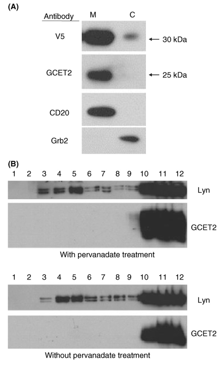
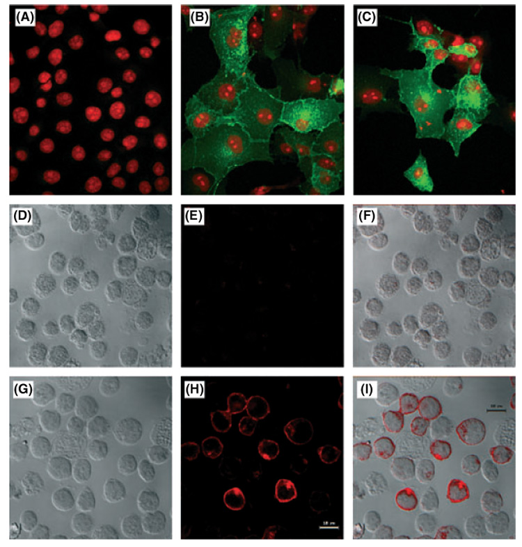
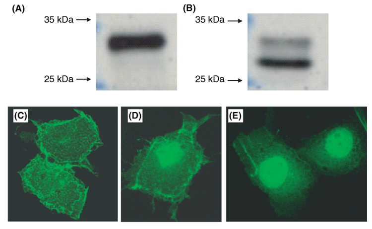
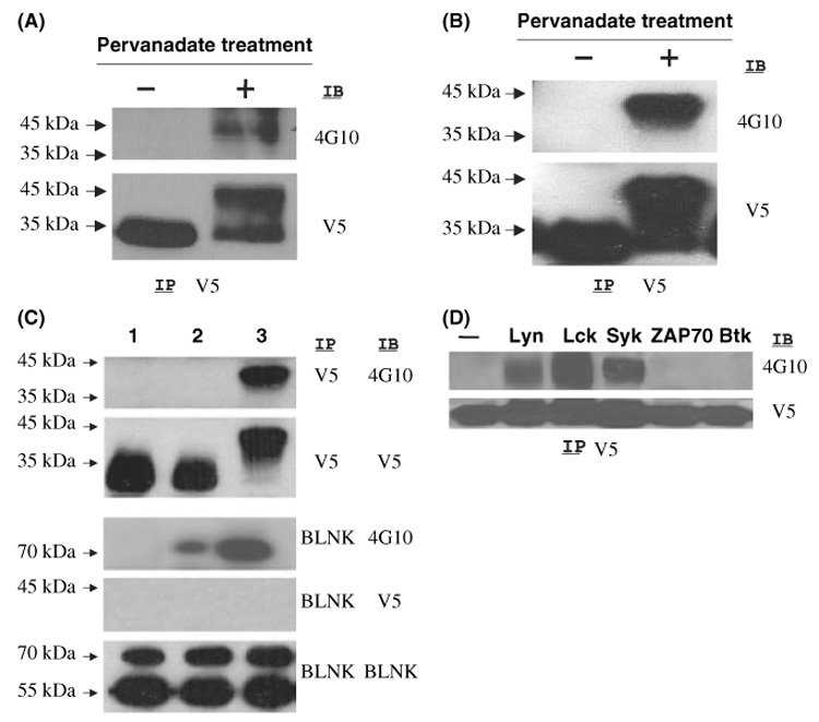
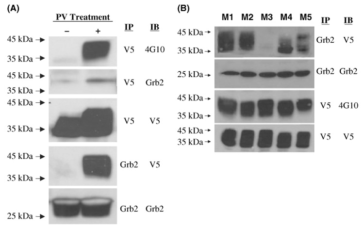
GCET2 (Germinal centre B-cell expressed transcript 2; also named HGAL) is a newly cloned gene that has been shown to be a useful marker for germinal centre (GC) B cells and GC B-cell derived malignancies, including follicular lymphomas and germinal centre B cell-like diffuse large B-cell lymphomas (GCB-DLBCLs), and is a useful prognosticator for DLBCLs. We report here the biochemical and biological properties of GCET2, which may help to determine its role in the GC reaction. GCET2 is constitutively localised in the plasma membrane but is excluded from lipid rafts. GCET2 does not have a transmembrane domain, and its membrane localisation is mediated by myristoylation and palmitoylation. GCET2 has five conserved putative tyrosine phosphorylation sites, and it can be phosphorylated following pervanadate treatment in B cells. By serially mutating the five tyrosines, the third and fourth tyrosines were found to be essential for GCET2 phosphorylation. GCET2 was phosphorylated when co-transfected into COS7 cells with protein tyrosine kinases (PTKs) LYN, LCK or SYK, and therefore it could be a substrate of these kinases in B cells. The third tyrosine site ((107)YENV) of GCET2 is a consensus GRB2 binding site, and GCET2 was found to associate with GRB2 through the third tyrosine following phosphorylation. Our data suggests that GCET2 may be an adaptor protein in GC B cells that transduces signals from GC B-cell membrane to the cytosol via its association with GRB2.


Figures

Fig 1
Fig 1
Alignment of germinal centre B-cell expressed transcript 2 (GCET2) with M17. M17 is a mouse germinal centre B-cell expressed gene and is 57% identical to GCET2. M17-L is an M17 isoform that was cloned by our laboratory from the mouse spleen and the mouse B-cell line, A20. The second exon is absent from the previously described shorter form of M17. The bolded and underlined sequences of GCET2 are the myristoylation site (¹MGNS) and the palmitoylation site (43CFC) respectively. The bolded sequences of GCET2 are the predicated tyrosine phosphorylation sites and all five are conserved in GCET2 and M17.
Fig 2
Fig 2
Germinal centre B-cell expressed transcript 2 (GCET2) is localised in the cell membrane but is excluded from the lipid rafts. (A) GCET2 is constitutively localised in the plasma membrane. The cell membrane (M) and the cytosol (C) were separated from DHL16 cells transduced with pMIG-GCET2. The upper panel was blotted using anti-V5 antibody to detect the exogenously expressed GCET2, and the second panel was blotted using anti-GCET2 antibody to detect the endogenous GCET2. CD20 and GRB2 were used as markers for the plasma membrane and cytosolic fractions, respectively. (B) GCET2 is excluded from the lipid rafts of cell membrane. GCET2 is not detected in the fractions 4–6 of the sucrose gradient, which represent the lipid rafts, before or after pervanadate treatment. LYN, known to be localised in the lipid rafts, was used as the positive control.
Fig 3
Fig 3
Localisation of germinal centre B-cell expressed transcript 2 (GCET2) in COS7 and B cells by confocal microscopy. (A) Localisation of GCET2 in COS7 cells. This figure shows the negative control. (B) Localisation of GCET2 in COS7 cells by confocal analysis. Wild-type GCET2 was mainly localised in the cell membrane with some distribution in the cytosol, probably in the Golgi apparatus and the membrane of cytosolic organelles. (C) Localisation of wild-type GCET2 in COS7 cells. (D) Localisation of GCET2 in DHL16 cells. DHL16 cells without pMIG-GCET2 transduction were used as negative control. Cells were stained using mouse anti-V5 monoclonal primary antibody and Texas red conjugated horse anti-mouse IgG secondary antibody. This picture is the Differential Interface Contrast (DIC) of control DHL16 B cells. (E) Fluorescence of control DHL16 cells. The cells were totally negative for red signals. (F) Combination (overlay) of G and H. (G) DIC of pMIG-GCET2 transduced B cell. (H) Red fluorescence of pMIG-GCET2 transduced B cells. Red signals were predominantly in the cell membrane and the Golgi area. (I) Combination (overlay) of J and K.
Fig 4
Fig 4
Germinal Centre B-cell Expressed Transcript 2 (GCET2) membrane localisation is mediated through myristoylation and palmitoylation. DHL16 cells were labelled with myristic acid [9, 10-³H (N)] or palmitic acid [9, 10-³H (N)]. GCET2 was both myristoylated (A) and palmitoylated (B). Myristoylation is a irreversible process that modifies one amino acid (Glycine-2) of GCET2, while palmitoylation is a reversible process that modifies two amino acids of GCET2 (Cysteine-46 and -48), and GCET2 in the lower molecular weight band (B) may represent the form that is not fully palmitoylated. (C) COS7 cells transfected with wild-type pcDNA3.1-GCET2 with V5 tag. COS7 cells were stained using FITC-labelled anti-V5 antibody. GCET2 protein is predominantly localised on the cell membrane; D. COS7 cells were transfected with GCET2 mutated in the myristoylation site. GCET2 had increased distribution in the nuclei; E. COS7 cells were transfected with GCET2 mutated in the myristoylation and the palmitoylation sites. GCET2 showed nuclear but not membranous distribution.
Fig 5
Fig 5
Germinal centre B-cell expressed transcript 2 (GCET2) is phosphorylated after PV Treatment of DHL16 Cells and Daudi Cells. (A) V5 tagged GCET2 was transiently transfected into DHL16 cells. Upon pervanadate treatment, phosphorylated GCET2 migrated to a band of higher molecular weight that reacted with 4G10. (B) GCET2 was phosphorylated in Daudi cells after pervanadate treatment. (C) BCR cross-linking cannot induce GCET2 phosphorylation in DHL16 cells. Both GCET2 and BLNK are phosphorylated upon pervanadate treatment, but BCR cross-linking can only induce BLNK phosphorylation. GCET2 cannot be co-precipitated with BLNK upon pervanadate treatment. Lane 1, DHL16 without treatment; 2, BCR cross-linking; 3, pervanadate treatment. (D) GCET2 is phosphorylated when co-transfected with LYN, LCK or SYK in COS7 cells. Anti-V5 antibody was used for immunoprecipitation (IP) followed by immunoblotting using the anti-phospho-tyrosine antibody, 4G10.
Fig 6
Fig 6
Constructs of germinal centre B-cell expressed transcript 2 (GCET2) mutants in the five conserved tyrosine sites. The tyrosines were mutated to phenylalanine. M1 to M5 represent the constructs with single mutations in the first to fifth tyrosine respectively, and M1+2 to M4+5 represent the constructs with mutation in the tyrosines indicated. W1 to W5 represent constructs with mutations of all tyrosines except the one indicated by ‘Y’. (B) Phosphorylation of GCET2 single mutants. Only the mutant in the third or fourth tyrosine has significantly decreased phosphorylation. (C) Phosphorylation of GCET2 combined mutants. All combined mutants that contain either the third or the fourth tyrosine have significantly decreased phosphorylation. (D) Phosphorylation of GCET2 truncated mutants and mutants on the lipid modification site. The truncated mutant that does not contain the fifth tyrosine (T5) has no decrease in tyrosine phosphorylation compared with the wild-type. Only very weak tyrosine phosphorylation can be detected in T45, the mutant that does not contain the fourth and the fifth tyrosines. The mutant on the myristoylation site (G2A) has no decrease of tyrosine phosphorylation. The phosphorylation of single GCET2 mutants is similar to the previous experiments, showing that only M3 and M4 have significant decreasing in phosphorylation levels. (E) GCET2 with one single tyrosine cannot be phosphorylated. No phosphorylation of W3, W4 or W5 was detected. Wild-type GCET2 co-transfected with LYN and SYK shows significantly increased phosphorylation after pervanadate treatment. IP, immunoprecipitation, IB, immunoblotting.
Fig 7
Fig 7
(A) Germinal centre B-cell expressed transcript 2 (GCET2) associates with GRB2 following pervanadate treatment. GCET2 is phosphorylated (first panel) and a weak signal of GRB2 is detected in the immunoprecipitates with V5 antibody (second panel). A strong signal of GCET2 is detected in the immunoprecipitates with GRB2 antibody (fourth panel). (B) The third tyrosine of GCET2 is important for its association with GRB2. Single mutants of GCET2 (M1 to M5) were transfected into COS7 cells together with LYN and SYK, and then anti-GRB2 antibody was used to precipitate GCET2. M3 has no association with GRB2. M1 and M2 had strong association to GRB2. M4 and M5 had slight decrease of association. IP, immunoprecipitation, IB, immunoblotting.

References

    1. Alizadeh AA, Eisen MB, Davis RE, Ma C, Lossos IS, Rosenwald A, Boldrick JC, Sabet H, Tran T, Yu X, Powell JI, Yang L, Marti GE, Moore T, Hudson J, Jr, Lu L, Lewis DB, Tibshirani R, Sherlock G, Chan WC, Greiner TC, Weisenburger DD, Armitage JO, Warnke R, Levy R, Wilson W, Grever MR, Byrd JC, Botstein D, Brown PO, Staudt LM. Distinct types of diffuse large B-cell lymphoma identified by gene expression profiling. Nature. 2000;403:503–511. - PubMed
    1. Allam A, Marshall AJ. Role of the adaptor proteins Bam32, TAPP1 and TAPP2 in lymphocyte activation. Immunology Letters. 2005;97:7–17. - PubMed
    1. Brdicka T, Imrich M, Angelisova P, Brdickova N, Horvath O, Spicka J, Hilgert I, Luskova P, Draber P, Novak P, Engels N, Wienands J, Simeoni L, Osterreicher J, Aguado E, Malissen M, Schraven B, Horejsi V. Non-T cell activation linker (NTAL): a transmembrane adaptor protein involved in immunoreceptor signaling. Journal of Experimental Medicine. 2002;196:1617–1626. - PMC - PubMed
    1. Cavenagh MM, Whitney JA, Carroll K, Zhang C, Boman AL, Rosenwald AG, Mellman I, Kahn RA. Intracellular distribution of Arf proteins in mammalian cells. Arf6 is uniquely localized to the plasma membrane. Journal of Biological Chemistry. 1996;271:21767–21774. - PubMed
    1. Chiu CW, Dalton M, Ishiai M, Kurosaki T, Chan AC. BLNK: molecular scaffolding through ‘cis’-mediated organization of signaling proteins. EMBO Journal. 2002;21:6461–6472. - PMC - PubMed

Publication types

MeSH terms