. 2007 Oct;122(2):210-21.
doi: 10.1111/j.1365-2567.2007.02630.x. Epub 2007 May 9.

Instruction of naive CD4+ T-cell fate to T-bet expression and T helper 1 development: roles of T-cell receptor-mediated signals

Affiliations

Instruction of naive CD4+ T-cell fate to T-bet expression and T helper 1 development: roles of T-cell receptor-mediated signals

Haruyuki Ariga et al. Immunology. 2007 Oct.

Erratum in

  • Immunology. 2007 Nov;122(3):454

Abstract

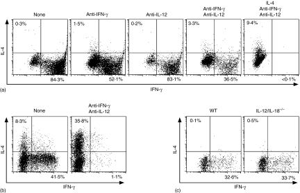
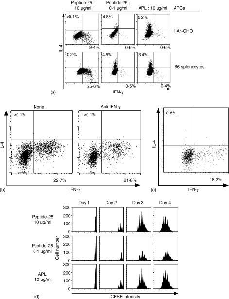
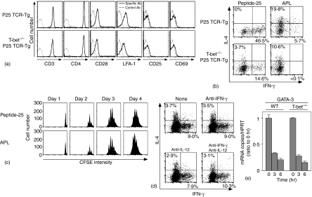
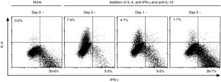
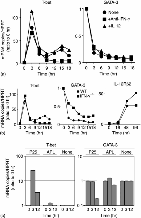
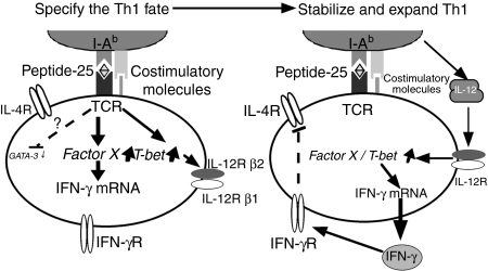
Using T-cell receptor (TCR) transgenic mice, we demonstrate that TCR stimulation of naive CD4(+) T cells induces transient T-bet expression, interleukin (IL)-12 receptor beta2 up-regulation, and GATA-3 down-regulation, which leads to T helper (Th)1 differentiation even when the cells are stimulated with peptide-loaded I-A(b)-transfected Chinese hamster ovary cells in the absence of interferon-gamma (IFN-gamma) and IL-12. Sustained IFN-gamma and IL-12 stimulation augments naive T-cell differentiation into Th1 cells. Intriguingly, a significant Th1 response is observed even when T-bet(-/-) naive CD4(+) T cells are stimulated through TCR in the absence of IFN-gamma or IL-12. Stimulation of naive CD4(+) T cells in the absence of IFN-gamma or IL-12 with altered peptide ligand, whose avidity to the TCR is lower than that of original peptide, fails to up-regulate transient T-bet expression, sustains GATA-3 expression, and induces differentiation into Th2 cells. These results support the notion that direct interaction between TCR and peptide-loaded antigen-presenting cells, even in the absence of T-bet expression and costimulatory signals, primarily determine the fate of naive CD4(+) T cells to Th1 cells.


Figures

Figure 1
Figure 1
Th1 differentiation of P25 TCR-Tg naive CD4+ T cells upon Peptide-25 stimulation is independent on IFN-γ, IL-12 and IL-18. (a) Naive CD4+ T cells from RAG-2–/– P25 TCR-Tg mice were stimulated in vitro with 10 µg/ml of Peptide-25 for 6 days in the presence of splenic APC. On day 0, some groups of culture received anti-IFN-γ, anti-IL-12, IL-4 or a combination of these, as depicted in (a). After the culture the primed cells were re-stimulated and IFN-γ- and IL-4-producing cells were assessed. Events shown are gated on live Vβ11+ cells. Results are presented for one of three experiments performed, with similar results in each experiment. (b) Naive CD4+ T cells from WT C57BL/6 were stimulated in vitro with 10 µg/ml of soluble anti-CD3 in the presence of splenic APC. On day 0, a group of culture received anti-IFN-γ and anti-IL12. After the culture the primed cells were re-stimulated and IFN-γ- and IL-4-producing cells were assessed. Events shown are gated on live CD4+ cells. Representative results of two separate experiments were displayed. (c) P25 TCR-Tg naive CD4+ T cells were stimulated in vitro with 10 µg/ml of Peptide-25 in the presence of splenic APC from WT or IL-12/IL-18–/– mice. After the culture the primed cells were re-stimulated and IFN-γ- and IL-4-producing cells were assessed. Events shown are gated on live Vβ11+ cells. Representative results of two separate experiments were displayed.
Figure 3
Figure 3
Th1 differentiation of P25 TCR-Tg naive CD4+ T cells can be induced in Peptide-25-loaded antigen-presenting cells lacking nominal costimulatory molecules. (a) P25 TCR-Tg naive CD4+ T cells were stimulated in vitro with 10 µg/ml or 0·1 µg/ml of Peptide-25 or 10 µg/ml of APL in the presence of I-Ab-CHO or splenic APC. After the culture the primed cells were re-stimulated and IFN-γ- and IL-4-producing cells were assessed. Events shown are gated on live Vβ11+ cells. Representative results of three separate experiments were displayed. (b) Naive CD4+ T cells from P25 TCR-Tg were stimulated in vitro with Peptide-25-loaded I-Ab-CHO in the presence or absence of anti-IFN-γ. After the culture the primed cells were re-stimulated and IFN-γ- and IL-4-producing cells were assessed. Events shown are gated on live Vβ11+ cells. Results are presented for one of three experiments performed, with similar results in each experiment. (c) Naive CD4+ T cells from STAT1–/– P25 TCR-Tg mice were stimulated in vitro with Peptide-25-loaded I-Ab-CHO. After the culture the primed cells were re-stimulated and IFN-γ- and IL-4-producing cells were assessed. Events shown are gated on live Vβ11+ cells. Results are presented for one of three experiments performed, with similar results in each experiment. (d) Naive CD4+ T cells from P25 TCR-Tg mice were labeled with 5 µm of CFSE and stimulated with 10 µg/ml or 0·1 µg/ml of Peptide-25 or 10 µg/ml of APL in the presence of splenic APC. On the days indicated, CD4+ T cells were harvested, stained for CD4 and Vβ11, and then analysed for dilution of CFSE intensity by FACSCalibur. Events shown are gated on live CD4+− Vβ11+ cells.
Figure 5
Figure 5
Induction of GATA-3 transcripts is down-regulated in Peptide-25-loaded I-Ab-CHO-stimulated T-bet–/– P25 TCR-Tg naive T cells. (a) Naive CD4+ T cells from P25 TCR-Tg or T-bet–/– P25 TCR-Tg mice were stained for CD3, CD4, CD28, LFA-1, CD25 and CD69. Events shown were gated on live cells. (b) Naive CD4+ T cells from P25 TCR-Tg or T-bet–/– P25 TCR-Tg mice were stimulated in vitro with 10 µg/ml of Peptide-25 in the presence of splenic APC under non-skewing conditions. After the culture the primed cells were re-stimulated and IFN-γ- and IL-4-producing cells were assessed. Events shown are gated on live Vβ11+ cells. Representative results of four separate experiments were displayed. (c) Naive CD4+ T cells from T-bet–/– P25 TCR-Tg mice were labelled with CFSE and stimulated with 10 µg/ml of Peptide-25 or 10 µg/ml of APL in the presence of splenic APC. On the days indicated, CD4+ T cells were harvested, stained for CD4 and Vβ11, and analysed for dilution of CFSE intensity by FACSCalibur. Events shown are gated on live CD4 ± Vβ11+ cells. (d) Naive CD4+ T cells from T-bet–/– P25 TCR-Tg mice were stimulated with Peptide-25-loaded I-Ab-CHO. On day 0, some groups of culture received anti-IFN-γ, anti-IL-12 or a combination of these, as depicted in (d). After the culture the primed cells were re-stimulated and IFN-γ- and IL-4-producing cells were assessed. Events shown are gated on live Vβ11+ cells. Results are presented for one of three experiments performed, with similar results in each experiment. (e) Naive CD4+ T cells from P25 TCR-Tg or T-bet–/– P25 TCR-Tg mice were stimulated for indicated periods of time (hr) with Peptide-25-loaded I-Ab-CHO. Cells were collected at the indicated time points and RNA was extracted. Quantitative real-time PCR was performed for assessing the mRNA expression of GATA-3 and HPRT. Each sample was normalized to HPRT. Data shown are ratios of test mRNA copies to mRNA copies of unstimulated cells. The values represent the mean and SD. Representative results of three separate experiments were displayed.
Figure 2
Figure 2
Commitment to Th1 differentiation of P25 TCR-Tg CD4+ T cells occurs within 3 days after stimulation with Peptide-25. P25 TCR-Tg naive CD4+ T cells were separated into four groups. Each group was stimulated with 10 µg/ml of Peptide-25 in the presence of splenic APC. IL-4, anti-IFN-γ and anti-IL-12 were added into each of three groups of culture on days 0, 1, and 3. After the culture the primed cells were re-stimulated and IFN-γ- and IL-4-producing cells were assessed. Events shown are gated on live Vβ11+ cells. Representative results of three separate experiments were displayed.
Figure 4
Figure 4
Kinetics of induction of T-bet, GATA-3 and IL-12Rβ2 transcripts in Peptide-25-loaded I-Ab-CHO-activated T cells. Naive CD4+ T cells were stimulated for indicated periods of time (hr) with Peptide-25- or APL-loaded I-Ab-CHO. Cells were collected at the indicated time points and RNA was extracted. Quantitative real-time PCR was performed for assessing the mRNA expression of T-bet, GATA-3 or IL-12Rβ2. Each sample was normalized to HPRT. Data shown are ratios of test mRNA copies to mRNA copies of unstimulated cells. The values represent the mean and SD. When error bars are not visible they are smaller than the symbol width. Results are presented for one of three experiments performed, with similar results in each experiment. (a) P25 TCR-Tg naive CD4+ T cells were stimulated with Peptide-25-loaded I-Ab-CHO alone or in the presence of anti-IFN-γ (3 µg/ml) or rIL-12 (10 ng/ml). (b) Naive CD4+ T cells either from WT P25 TCR-Tg mice or IFN-γ–/– P25 TCR-Tg mice were stimulated with Peptide-25-loaded I-Ab-CHO. (c) P25 TCR-Tg naive CD4+ T cells were stimulated with Peptide-25-loaded I-Ab-CHO or APL-loaded I-Ab-CHO. As a control, cells were incubated without any stimulus.
Figure 6
Figure 6
The pathway of Th1 development illustrates how external TCR signals play a role differently from IFN-γ and IL-12, and how these pathways are intrinsically ordered. (Left-hand panel) Antigen receptor ligation enables T-bet to specify the Th1 fate, including T-bet auto-induction, IL-12Rβ2 induction, and primary remodelling of the ifn-γ locus. (Right-hand panel) The actions of T-bet then enable IFN-γ and probably IL-12 to signal for survival and growth of Th1 cells, and interact for secondary enhancement of IFN-γ gene expression.

Similar articles

Cited by

References

    1. Mosmann TR, Cherwinski H, Bond MW, Giedlin MA, Coffman RL. Two types of murine helper T cell clone. I. Definition according to profiles of lymphokine activities and secreted proteins. J Immunol. 1986;136:2348–57. - PubMed
    1. Abbas AK, Murphy KM, Sher A. Functional diversity of helper T lymphocytes. Nature. 1996;383:787–93. - PubMed
    1. O'Garra A. Cytokines induce the development of functionally heterogeneous T helper cell subsets. Immunity. 1998;8:275–83. - PubMed
    1. Murphy KM, Ouyang W, Farrar JD, Yang J, Ranganath S, Asnagli H, Afkarian M, Murphy TL. Signaling and transcription in T helper development. Annu Rev Immunol. 2000;18:451–94. - PubMed
    1. O'Shea JJ, Paul WE. Regulation of TH1 differentiation – controlling the controllers. Nat Immunol. 2002;3:506–8. - PubMed

Publication types

MeSH terms

LinkOut - more resources