. 2007 Jul;85(1):130-41.
doi: 10.1016/j.exer.2007.03.009. Epub 2007 Mar 31.

Injury and nucleotides induce phosphorylation of epidermal growth factor receptor: MMP and HB-EGF dependent pathway

Affiliations

Injury and nucleotides induce phosphorylation of epidermal growth factor receptor: MMP and HB-EGF dependent pathway

Ilene Boucher et al. Exp Eye Res. 2007 Jul.

Abstract

The early events that occur rapidly after injury trigger signal cascades that are essential for proper wound closure of corneal epithelial cells. We hypothesize that injury releases ATP, which stimulates purinergic receptors and elicits the phosphorylation of epidermal growth factor receptor (EGFR) tyrosine residues and subsequent cell migration by a MMP and HB-EGF dependent pathway. We demonstrated that the inhibition of purinergic receptors with the antagonist, Reactive Blue 2, abrogated the phosphorylation of EGFR and ERK. Pre-incubation of cells with the EGFR kinase inhibitor, AG1478, and subsequent stimulation by injury or ATP resulted in a decrease in phosphorylation of EGFR and migration. Furthermore, downregulation of EGFR by siRNA, inhibited the EGF-induced intracellular Ca(2+) wave. However, the response to injury and ATP was retained indicating the presence of two signaling pathways. Inhibition with either CRM197 or TIMP-3 decreased injury and nucleotide-induced phosphorylation of both EGFR and ERK. Incubation in the presence of a functional blocking antibody to HB-EGF also resulted in a decrease in the phosphorylation of EGFR. In addition, cell migration was inhibited by CRM197 and rescued when cells were incubated with HB-EGF. We showed that injury-induced phosphorylation of specific tyrosine residues and found that a similar pattern of phosphorylation was induced by trinucleotides. These studies indicate that injury-induced purinergic receptor activation leads to phosphorylation of EGFR, ERK and migration.


Figures

Fig. 1
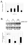
Fig. 1. Role of ATP in injury induced response
A. ATP bioluminescence assay of injured cells compared to control cells. N=6 independent cultures. B. Cells were incubated in the presence or absence of Reactive Blue 2 (RB-2) (100 μM) and injured (W) or stimulated with ATP and incubated for 5 min. Untreated cells (medium change +/− RB-2) were used as control. Lysates of equivalent protein concentration were resolved in SDS-PAGE and immunoblotted with antibodies directed against phosphotyrosine 1173 (pEGFR), pan-EGFR, total and phosphoERK. N=3 independent experiments. C. Cells were preincubated with AG1478, and subjected to injury, ATP, or EGF for 5 min. Untreated cells (medium change) were used as control. Lysates of equivalent protein concentration were immunoprecipitated with EGFR and immunoblotted with PY20 or with EGFR. In parallel experiments equivalent amounts of protein were resolved on SDS-PAGE and immunoblotted with antibodies directed against total and phosphoERK. N=5 independent experiments. D. Cells were incubated in the presence or absence of AG1478 (10 nM) and stimulated with ATP, EGF or binding buffer (−). Transwell migrations were performed for 8 hr at 37°C. The migrated cells were stained with propidium iodide, counted in six randomly chosen fields, averaged and normalized to unstimulated control. N=3 independent experiments.
Fig. 2
Fig. 2. siRNA to EGFR does not reduce Ca2+ mobilization but reduces pERK
A. Lysates from cells transfected with siRNA to EGFR were probed for EGFR. Equivalent amounts of protein were resolved in SDS-PAGE and blotted with a pan-EGFR antibody. B. Cells transfected with the siRNA and control sequences were incubated in fluo-3/AM and imaged using live cell confocal microscopy. Cells were stimulated with EGF, ATP or subjected to injury (W). The data is presented as relative fluorescence. ** t-test (p<0.001). C. The cells imaged were lysed and equivalent protein was immunoblotted onto membranes and probed with antibodies to pERK and ERK. * t-test (p<0.05). D. PAE cells (lacking or stably transfected with EGFR) were stimulated with injury (W), ATP or EGF for 5 min and lysates were probed as described above. (N.D -not detected). Phosphorylation was normalized to total ERK and a representative blot was graphed as fold change over control. N= 3 independent experiments for A, B, C and D.
Fig. 3
Fig. 3. Injury induced phosphorylation of EGFR is mediated by HB-EGF
A. Cells were incubated in the presence or absence of the functional blocking antibody to HB-EGF (10 μg/ml) and subjected to control medium, injury (W), or HB-EGF (10 nM). Equivalent amounts of protein were immunoprecipitated with pan-EGFR antibody, resolved on SDS-PAGE and immunoblotted with PY20. Phosphorylation was normalized to EGFR and the graph is presented as relative phosphorylation. B. Cell migration to HB-EGF is concentration dependent. Cells were stimulated with HB-EGF, EGF or binding buffer (−). Transwell migrations were performed for 8 hr at 37°C. The migrated cells were stained with propidium iodide, counted in six randomly chosen fields, averaged and normalized to unstimulated control. N=3 independent experiments.
Fig. 4
Fig. 4. Injury and ATP induced phosphorylation of EGFR is mediated by cleavage of the HB-EGF
A. Cells were incubated in the presence or absence of CRM197 (40 μg/ml) and then subjected to control medium, injury (W), ATP or EGF. Equivalent amounts of protein were immunoprecipitated with pan-EGFR antibody, resolved in SDS-PAGE and immunoblotted with either PY20 or EGFR. B. Cells were incubated in the presence or absence of TIMP-3 (50 ng/ml) and then subjected to control medium, injury (W), ATP or EGF. Equivalent amounts of protein were immunoprecipitated with pan-EGFR, resolved on SDS-PAGE and blotted with either PY20 or EGFR. Densitometric analysis was performed and graphs are presented as normalized to EGFR for each condition in A. and B. C. Cells were incubated in the presence or absence of CRM197 (40 μg/ml) and subjected to control medium, injury (W), ATP or EGF. Lysates were harvested after 5 min. Equivalent amounts of protein were resolved in SDS-PAGE and immunoblotted with anti-ERK. Membranes were probed with antibodies directed against phospho and total ERK. D. Cells were incubated in the presence or absence of TIMP-3 (50 ng/ml) and subjected to control medium, injury (W), ATP or EGF as described above. Equivalent amounts of protein were resolved in SDS-PAGE and blotted with antibodies directed against pERK and total ERK. Graphs are normalized to total ERK for C. and D. N=3 independent experiments for A, B, C, and D.
Fig. 5
Fig. 5. CRM197 inhibits wound repair
Confluent cells in 8 well chamber slides were incubated overnight in medium lacking growth factors. Cells were pre-incubated in the presence or absence of CRM197 (40 μg/ml) or CRM197 (40 μg/ml) and HB-EGF (10 nM), placed on a heated microscope stage, wounded and incubated in an environmental chamber at 37°C and 5% CO2 for 20 hours. The wounds were demarcated and contiguous regions were tiled and imaged every 20 min. A. Migration time course of a representative run. B. Percent closure at 20 hours. *p<0.01 Significance was determined by a one-way ANOVA followed by Tukey's post hoc test. N=3 independent experiments.
Fig. 6
Fig. 6. Injury and nucleotides elicit phosphorylation of EGFR
A. Cells were cultured and subjected to control (medium change), injury (W), ATP, or EGF. Equivalent amounts of protein were resolved on SDS-PAGE and immunoblotted with polyclonal antibodies directed against specific phosphotyrosine sites on EGFR (845, 1068, 1086, 1148 and 1173). Blots were probed also for EGFR. Densitometric analysis was performed and graphs are presented as fold change over control. B. Immunohistochemical analysis. Cells were cultured, a linear wound was made and cells were washed and fixed at five min. In fifty percent of the cultures, cells were preincubated for one hour with serum free medium containing TIMP-3 while the other half were incubated in serum free medium. Cells were permeabilized, blocked and probed with polyclonal antibodies directed against the phosphotyrosine sites and detected with Alexa 488-conjugated IgG. Fluorescent and phase images were taken and merged. Negative control lacks primary antibody. Confocal micrographs represent single optical sections of 3μm. Micrographs are representative of two independent experiments (scale bar equals 100 μm). C. Differential phosphorylation of EGFR in response to ATP, UTP, ADP, UDP compared to control. Equivalent amounts of protein were resolved in SDS-PAGE and blotted with antibodies directed against phosphotyrosine specific sites on EGFR (845, 1068, 1086, 1148 and 1173). Blots were probed also for total EGFR. Densitometric analysis was performed and graphs are presented as fold change over control. N- 3 independent experiments.
Fig.7
Fig.7. Schematic of cellular response to injury and trinucleotides

Similar articles

Cited by

References

    1. Abbracchio MP, Burnstock G. Purinergic signalling: pathophysiological roles. Jpn J Pharmacol. 1998;78:113–45. - PubMed
    1. Andreev J, Galisteo ML, Kranenburg O, Logan SK, Chiu ES, Okigaki M, Cary LA, Moolenaar WH, Schlessinger J. Src and Pyk2 Mediate G-protein-coupled Receptor Activation of Epidermal Growth Factor Receptor (EGFR) but are not Required for Coupling to the Mitogen-activated Protein (MAP) Kinase Signaling Cascade. J Biol Chem. 2001;276:20130–20135. - PubMed
    1. Araki-Sasaki K, Ohashi Y, Sasabe T, Hayashi K, Watanabe H, Tano Y, Handa H. An SV40-immortalized human corneal epithelial cell line and its characterization. Invest Ophthalmol Vis Sci. 1995;36:614–621. - PubMed
    1. Block ER, Matela A, SundarRaj N, Iszkula ER, Klarlund JK. Wounding induces motility in sheets of corneal epithelial cells through loss of spatial constraints: role of heparin-binding epidermal growth factor-like growth factor signaling. J Biol Chem. 2004;279:24307–24312. - PubMed
    1. Burnstock G. The past, present and future of purine nucleotides as signalling molecules. Neuropharmacology. 1997;36:1127–39. - PubMed

Publication types

MeSH terms