. 2007 Jun 8;146(4):1593-605.
doi: 10.1016/j.neuroscience.2007.04.005. Epub 2007 May 9.

Dendritic distributions of dopamine D1 receptors in the rat nucleus accumbens are synergistically affected by startle-evoking auditory stimulation and apomorphine

Affiliations

Dendritic distributions of dopamine D1 receptors in the rat nucleus accumbens are synergistically affected by startle-evoking auditory stimulation and apomorphine

Y Hara et al. Neuroscience. .

Abstract

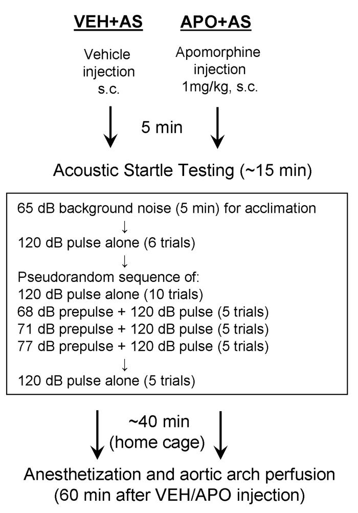
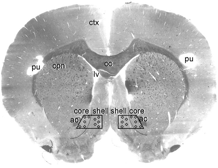
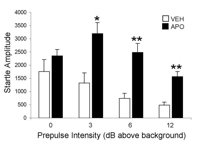
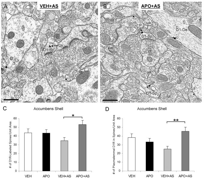
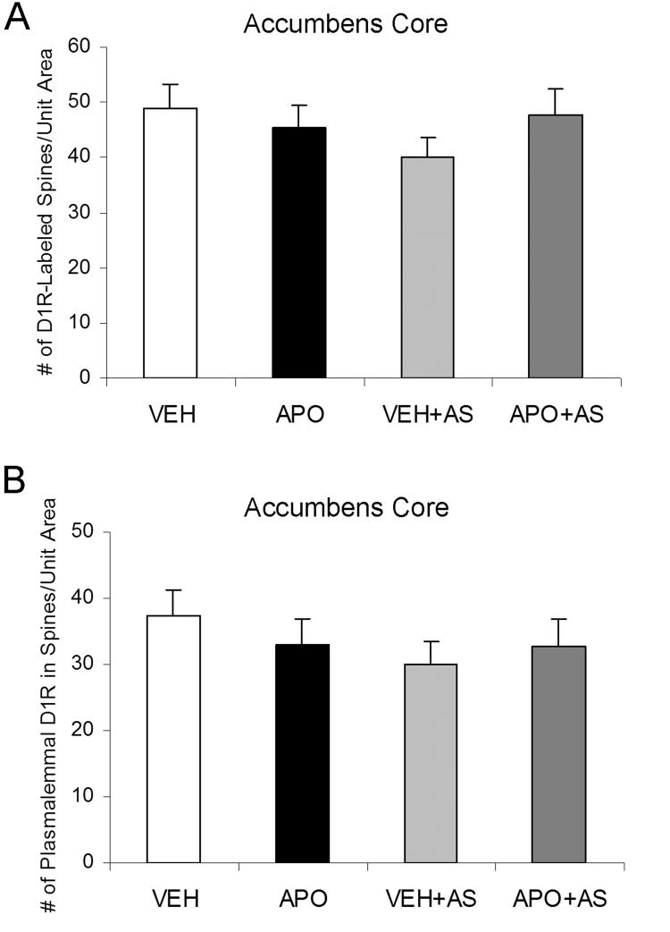
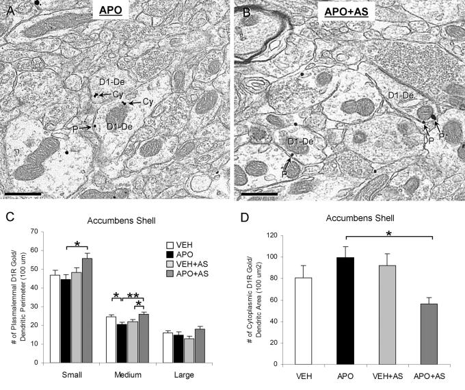
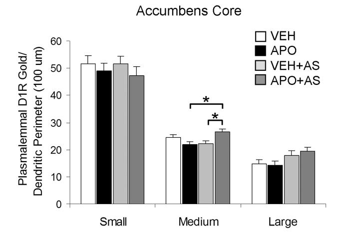
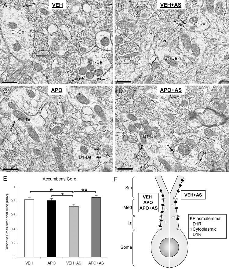
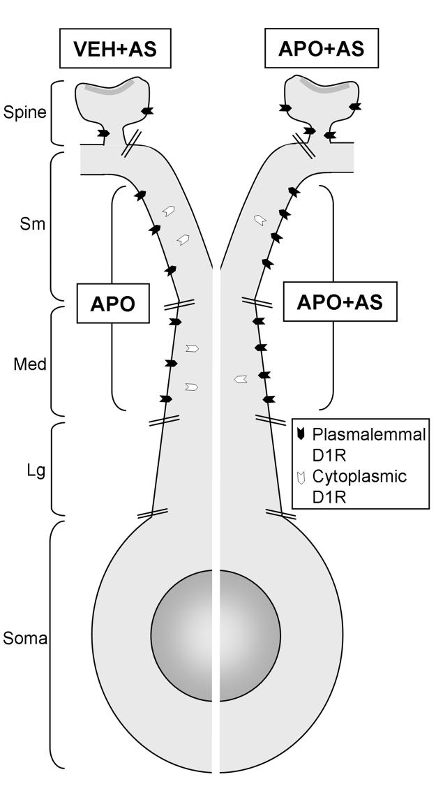
Prepulse inhibition of the startle response to auditory stimulation (AS) is a measure of sensorimotor gating that is disrupted by the dopamine D1/D2 receptor agonist, apomorphine. The apomorphine effect on prepulse inhibition is ascribed in part to altered synaptic transmission in the limbic-associated shell and motor-associated core subregions of the nucleus accumbens (Acb). We used electron microscopic immunolabeling of dopamine D1 receptors (D1Rs) in the Acb shell and core to test the hypothesis that region-specific redistribution of D1Rs is a short-term consequence of AS and/or apomorphine administration. Thus, comparisons were made in the Acb of rats killed 1 h after receiving a single s.c. injection of vehicle (VEH) or apomorphine (APO) alone or in combination with startle-evoking AS (VEH+AS, APO+AS). In both regions of all animals, the D1R immunoreactivity was present in somata and large, as well as small, presumably more distal dendrites and dendritic spines. In the Acb shell, compared with the VEH+AS group, the APO+AS group had more spines containing D1R immunogold particles, and these particles were more prevalent on the plasma membranes. This suggests movement of D1Rs from distal dendrites to the plasma membrane of dendritic spines. Small- and medium-sized dendrites also showed a higher plasmalemmal density of D1R in the Acb shell of the APO+AS group compared with the APO group. In the Acb core, the APO+AS group had a higher plasmalemmal density of D1R in medium-sized dendrites compared with the APO or VEH+AS group. Also in the Acb core, D1R-labeled dendrites were significantly smaller in the VEH+AS group compared with all other groups. These results suggest that alerting stimuli and apomorphine synergistically affect distributions of D1R in Acb shell and core. Thus adaptations in D1R distribution may contribute to sensorimotor gating deficits that can be induced acutely by apomorphine or develop over time in schizophrenia.


Figures

Figure 1
Figure 1
A schematic diagram of the experimental paradigm for VEH+AS and APO+AS groups. Five minutes after the rats received either a vehicle or apomorphine subcutaneous injection, they were introduced to the acoustic startle apparatus. The startle testing started with 5 minutes of 65 dB background noise to allow the rats to acclimate to the environment. Then they were given six 120 dB pulse alone trials followed by a pseudorandom sequence of pulse alone trials and prepulse+pulse trials. The session concluded with an additional five pulse alone trials. After the completion of acoustic startle testing, the VEH+AS and APO+AS rats were returned to their home cage for approximately 40 minutes until the perfusion (60 minutes after the vehicle/apomorphine injection). The VEH and APO groups also received a single subcutaneous injection of vehicle and apomorphine, respectively, but did not undergo acoustic startle testing and remained in their home cage for 60 minutes until the perfusion (not shown).
Figure 2
Figure 2
A light micrograph of a flat-embedded accumbens vibratome section labeled with D1R. The trapezoids indicate the locations from which the ultrathin sections were collected for electron microscopic image analysis. Each trapezoid included both the Acb shell and core, directly medial to the anterior commissure (ac) at the level 1.6 mm anterior to Bregma (Paxinos and Watson, 1986). Microscopic images were taken from 18-20 grid squares within the two Acb trapezoids per rat, illustrated by the small squares. None of these grid squares were adjacent to one another in order to collect a representative sample from a vast area of the medial Acb shell and core. Holes were punched (pu) at different locations within the cortex (ctx) to distinguish between rats in different experimental groups. cc, corpus callosum; cpn, caudate-putamen nucleus; lv, lateral ventricle.
Figure 3
Figure 3
A bar graph showing the maximal startle amplitude in vehicle (VEH)- and apomorphine (APO)- injected rats in response to 120 dB pulse alone (0) and prepulse (3, 6, 12db above background) plus 120 dB pulse. A two-way ANOVA followed by Tukey's post hoc test was used for statistical comparisons between the vehicle and apomorphine-injected rats. *p<0.05, **p<0.01, n=3 rats/experimental group
Figure 4
Figure 4
Apomorphine effects on D1R immunogold distributions in dendritic spines of the Acb shell in rats exposed to acoustic startle. (A, B) Electron micrographs showing spine heads (D1-Sp) with D1R immunogold particles in the cytoplasm (Cy) and on the plasma membrane (P) in VEH+AS group (A) and APO+AS group (B), respectively. The spines receive asymmetric excitatory-type synapse from unlabeled terminals (ut). In the APO+AS group, D1R immunogold particles are also present on the plasmalemma of a spine neck (D1-Sp2) and on the plasma membrane of a dendrite (D1-De). The plasmalemmal D1R immunogold particles are located near unlabeled small axonal profiles (ua). (C, D) Bar graphs showing the number of D1R-labeled spines (C) and the number of D1R gold particles on the plasma membrane of spines (D) per 4100 μm2 of Acb shell tissue. A one-way ANOVA followed by Tukey's post hoc test was used for statistical comparisons between the following groups: vehicle-injected (VEH), apomorphine-injected (APO), vehicle injection followed by auditory stimulation (VEH+AS) and apomorphine injection followed by auditory stimulation (APO+AS). *p<0.05, **p<0.01. n=3 rats/each of the four experimental groups
Figure 5
Figure 5
Bar graphs showing the between-group similarities in the numbers of D1R-labeled spines (A) and the numbers of D1R gold particles on the plasma membrane of spines (B) per 4100 μm2 of Acb core tissue. A one-way ANOVA followed by Tukey's post hoc test was used for statistical comparisons between the following groups: vehicle-injected (VEH), apomorphine-injected (APO), vehicle injection followed by auditory stimulation (VEH+AS) and apomorphine injection followed by auditory stimulation (APO+AS). No statistically significant differences were observed between any of the groups. n=3 rats/each of the four experimental groups
Figure 6
Figure 6
Size- and treatment-dependent differences in plasmalemmal and cytoplasmic D1R labeling in the Acb shell dendrites. (A, B) Electron micrographs showing differences in D1R immunogold distributions in small-medium dendrites (D1-De) between the APO (A) and APO+AS (B) groups. In the APO group, one dendrite contains D1R immunogold particles only in the cytoplasm (Cy) and the other dendrite contains both cytoplasmic and plasmalemmal (P) D1R gold particles. In the APO+AS group, the D1R immunogold particles in both dendrites are located on the plasma membrane (P). (C) A bar graph showing the number of plasmalemmal D1R immunogold particles per unit perimeter length (100 μm) in each experimental group and each size of Acb shell dendrites. Cluster analysis by dendritic diameter was performed to statistically divide the D1R-labeled dendrites into small, medium and large subcategories. A one-way ANOVA followed by Tukey's post hoc test was used for statistical comparisons between the following groups: vehicle-injected (VEH), apomorphine-injected (APO), vehicle injection followed by auditory stimulation (VEH+AS) and apomorphine injection followed by auditory stimulation (APO+AS). *p<0.05, **p<0.01, n=3 rats/each of the four experimental groups. (D) A bar graph showing the number of cytoplasmic D1R immunogold particles per unit dendritic cross-sectional area (100 um2) in the four experimental groups. A one-way ANOVA followed by Tukey's post hoc test was used for statistical comparisons between the groups. *p<0.05, n=3 rats/each of the four experimental groups
Figure 7
Figure 7
A bar graph showing an effect of apomorphine and auditory stimulation in the number of plasmalemmal D1R immunogold particles per unit perimeter length (100 μm), exclusively in medium-sized dendrites of the Acb core. Cluster analysis by dendritic diameter was performed to statistically divide the D1R-labeled dendrites into small, medium and large subcategories. A one-way ANOVA followed by Tukey's post hoc test was used for statistical comparisons between the following groups: vehicle-injected (VEH), apomorphine-injected (APO), vehicle injection followed by auditory stimulation (VEH+AS) and apomorphine injection followed by auditory stimulation (APO+AS). *p<0.05, n=3 rats/each of the four experimental groups
Figure 8
Figure 8
Targeting D1Rs to smaller dendrites of Acb core in rats exposed to vehicle injection and auditory stimulation (VEH+AS) compared with rats receiving a vehicle injection alone (VEH), an apomorphine injection alone (APO) or an apomorphine injection followed by auditory stimulation (APO+AS). (A-D) Electron micrographs showing D1R-labeled dendrites (D1-De) in each of the four groups (VEH, APO, VEH+AS, APO+AS). The D1R immunogold particles are located on the plasma membrane (P) of the dendrites in all groups. The D1R-labeled dendrites in the VEH+AS group are smaller in size (B) than those in all other groups (A, C, D). (E) A bar graph showing the average cross-sectional area (μm2) of Acb core D1R-labeled dendrites in each experimental group. A one-way ANOVA followed by Tukey's post hoc test was used for statistical comparisons between the following groups: VEH, APO, VEH+AS, and APO+AS. *p<0.05, **p<0.01; n=3 rats/each of the four experimental groups. (F) A schematic diagram showing the D1Rs targeted to smaller, presumably more distal dendrites in the VEH+AS group compared to VEH, APO and APO+AS groups in the Acb core. Sm, small dendrite; Med, medium dendrite; Lg, large dendrite.
Figure 9
Figure 9
A schematic diagram showing the soma and dendrites of a spiny neuron in Acb shell. The relative plasmalemmal/cytoplasmic densities of D1R gold particles in dendritic compartments (dendritic spine, small (Sm)-, medium (Med)-, and large (Lg)-sized dendrites) are shown. The neuron is artificially divided (white line) so as to compare experimental groups. These groups received a vehicle injection followed by auditory stimulation (VEH+AS), an apomorphine injection alone (APO), or an apomorphine injection followed by auditory stimulation (APO+AS). In spines, the APO+AS group has significantly more plasmalemmal D1Rs compared to the VEH+AS group. In small and medium dendrites, the APO+AS group has significantly more D1Rs on the plasma membrane compared to the APO group. The greater density of plasmalemmal D1R particles in the APO+AS group is paralleled by a decrease in cytoplasmic D1Rs, suggesting surface trafficking from a cytoplasmic reserve pool.

Similar articles

Cited by

References

    1. Bakshi VP, Tricklebank M, Neijt HC, Lehmann-Masten V, Geyer MA. Disruption of prepulse inhibition and increases in locomotor activity by competitive N-methyl-D-aspartate receptor antagonists in rats. J Pharmacol Exp Ther. 1999;288:643–652. - PubMed
    1. Braff DL, Grillon C, Geyer MA. Gating and habituation of the startle reflex in schizophrenic patients. Arch Gen Psychiatry. 1992;49:206–215. - PubMed
    1. Brog JS, Salyapongse A, Deutch AY, Zahm DS. The patterns of afferent innervation of the core and shell in the “accumbens” part of the rat ventral striatum: immunohistochemical detection of retrogradely transported fluoro-gold. J Comp Neurol. 1993;338:255–278. - PubMed
    1. Carey MP, Diewald LM, Esposito FJ, Pellicano MP, Gironi Carnevale UA, Sergeant JA, Papa M, Sadile AG. Differential distribution, affinity and plasticity of dopamine D-1 and D-2 receptors in the target sites of the mesolimbic system in an animal model of ADHD. Behav Brain Res. 1998;94:173–185. - PubMed
    1. Chan J, Aoki C, Pickel VM. Optimization of differential immunogold-silver and peroxidase labeling with maintenance of ultrastructure in brain sections before plastic embedding. J Neurosci Methods. 1990;33:113–127. - PMC - PubMed

Publication types

MeSH terms