

Abnormal cortical folding patterns within Broca's area in schizophrenia: evidence from structural MRI
- PMID: 17490861
- PMCID: PMC2034662
- DOI: 10.1016/j.schres.2007.03.031
Abnormal cortical folding patterns within Broca's area in schizophrenia: evidence from structural MRI
Abstract
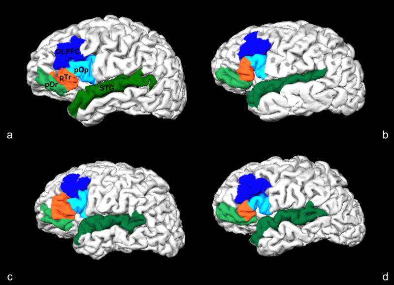
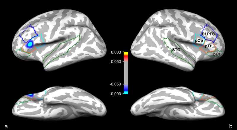
We compared cortical folding patterns between patients with schizophrenia and demographically-matched healthy controls in prefrontal and temporal regions of interest. Using the Freesurfer (http://surfer.nmr.mgh.harvard.edu) cortical surface-based reconstruction methodology, we indirectly ascertained cortical displacement and convolution, together, by measuring the degree of metric distortion required to optimally register cortical folding patterns to an average template. An area within the pars triangularis of the left inferior frontal gyrus (Broca's area) showed significantly reduced metric distortion in the patient group relative to the control group (p=0.0352). We discuss these findings in relation to the neurodevelopmental hypothesis and language dysfunction in schizophrenia.
Figures
Similar articles
-
Abnormal angular gyrus asymmetry in schizophrenia.Am J Psychiatry. 2000 Mar;157(3):428-37. doi: 10.1176/appi.ajp.157.3.428. Am J Psychiatry. 2000. PMID: 10698820 Free PMC article.
-
Automated ROI-based brain parcellation analysis of frontal and temporal brain volumes in schizophrenia.Psychiatry Res. 2006 Oct 30;147(2-3):153-61. doi: 10.1016/j.pscychresns.2006.04.007. Epub 2006 Sep 1. Psychiatry Res. 2006. PMID: 16949259
-
When Broca goes uninformed: reduced information flow to Broca's area in schizophrenia patients with auditory hallucinations.Schizophr Bull. 2013 Sep;39(5):1087-95. doi: 10.1093/schbul/sbs107. Epub 2012 Oct 15. Schizophr Bull. 2013. PMID: 23070537 Free PMC article.
-
Familial and developmental abnormalities of front lobe function and neurochemistry in schizophrenia.J Psychopharmacol. 1997;11(2):133-42. doi: 10.1177/026988119701100206. J Psychopharmacol. 1997. PMID: 9254279 Review.
-
Beyond a single area: motor control and language within a neural architecture encompassing Broca's area.Cortex. 2006 May;42(4):503-6. doi: 10.1016/s0010-9452(08)70387-3. Cortex. 2006. PMID: 16881259 Review.
Cited by
-
Chaos analysis of the cortical boundary for the recognition of psychosis.J Psychiatry Neurosci. 2023 Apr 25;48(2):E135-E142. doi: 10.1503/jpn.220160. Print 2023 Mar-Apr. J Psychiatry Neurosci. 2023. PMID: 37185319 Free PMC article.
-
Elevated peripheral cytokines characterize a subgroup of people with schizophrenia displaying poor verbal fluency and reduced Broca's area volume.Mol Psychiatry. 2016 Aug;21(8):1090-8. doi: 10.1038/mp.2015.90. Epub 2015 Jul 21. Mol Psychiatry. 2016. PMID: 26194183 Free PMC article.
-
Verbal fluency deficits and altered lateralization of language brain areas in individuals genetically predisposed to schizophrenia.Schizophr Res. 2009 Dec;115(2-3):202-8. doi: 10.1016/j.schres.2009.09.033. Epub 2009 Oct 17. Schizophr Res. 2009. PMID: 19840895 Free PMC article.
-
Gyral folding pattern analysis via surface profiling.Neuroimage. 2010 Oct 1;52(4):1202-14. doi: 10.1016/j.neuroimage.2010.04.263. Epub 2010 May 26. Neuroimage. 2010. PMID: 20472071 Free PMC article.
-
Altered language network activity in young people at familial high-risk for schizophrenia.Schizophr Res. 2013 Dec;151(1-3):229-37. doi: 10.1016/j.schres.2013.09.023. Epub 2013 Oct 28. Schizophr Res. 2013. PMID: 24176576 Free PMC article.
References
-
- American Psychiatric Association . DSM-IV: Diagnostic and Statistical Manual of Mental Disorders. 4th Rev ed. American Psychiatric Press; Washington, DC: 1990.
-
- Amunts K, Schleicher A, Ditterich A, Zilles K. Broca's region: cytoarchitectonic asymmetry and developmental changes. J Comp Neurol. 2003;465:72–89. - PubMed
-
- Andreasen NC. Thought, language and communication disorders: I. Clinical assessment, definition of terms, and evaluation of their reliability. Arch Gen Psychiatry. 1979;36:1315–1321. - PubMed
-
- Barde LH, Thompson-Schill SL. Models of functional organization of the lateral prefrontal cortex in verbal working memory: evidence in favor of the process model. J Cogn Neurosci. 2002;14:1054–1063. - PubMed
-
- Bartzokis G, Beckson M, Lu PH, Nuechterlein KH, Edwards N, Mintz J. Age-related changes in frontal and temporal lobe volumes in men: a magnetic resonance imaging study. Arch Gen Psychiatry. 2001;58:461–465. - PubMed
Publication types
MeSH terms
Grants and funding
- U24 RR021382/RR/NCRR NIH HHS/United States
- P41-RR14075/RR/NCRR NIH HHS/United States
- T32 CA009502/CA/NCI NIH HHS/United States
- R01 EB001550/EB/NIBIB NIH HHS/United States
- U54 EB005149/EB/NIBIB NIH HHS/United States
- P41 RR014075/RR/NCRR NIH HHS/United States
- R01 NS052585/NS/NINDS NIH HHS/United States
- R01 MH067720/MH/NIMH NIH HHS/United States
- R01 RR16594-01A1/RR/NCRR NIH HHS/United States
- R01 RR016594/RR/NCRR NIH HHS/United States
- R01 MH071635/MH/NIMH NIH HHS/United States
- 5T32 CA09502/CA/NCI NIH HHS/United States
- R01 NS052585-01/NS/NINDS NIH HHS/United States
LinkOut - more resources
Full Text Sources
Medical
Miscellaneous
